. 2011:105:3-24.
doi: 10.1016/B978-0-12-381320-6.00001-1.

Zebrafish models of germ cell tumor

Affiliations

Zebrafish models of germ cell tumor

Joanie C Neumann et al. Methods Cell Biol. 2011.

Abstract

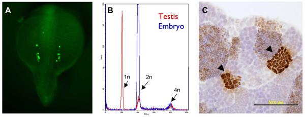
Germ cell tumors are neoplasms arising from pluripotent germ cells. In humans, these tumors occur in infants, children and young adults. The tumors display a wide range of histologic differentiation states which exhibit different clinical behaviors. Information about the molecular basis of germ cell tumors, and representative animal models of these neoplasms, are lacking. Germline development in zebrafish and humans is broadly conserved, making the fish a useful model to probe the connections between germ cell development and tumorigenesis. Here, we provide an overview of germline development and a brief review of germ cell tumor biology in humans and zebrafish. We also outline some methods for studying the zebrafish germline.


Figures

Figure 1
Figure 1. Histology of zebrafish germ cell tumors
Hematoxylin & eosin stained sections from formalin-fixed, paraffin-embedded zebrafish testes. A, B: normal testis. The testis consists of cysts or lobules of spermatogenic cells surrounded by a basement membrane and somatic cells. Small clusters of spermatogonia are seen adjacent to the basement membrane (arrowheads). Successive stages of differentiation are also evident, including primary and secondary spermatocytes, spermatids and mature spermatozoa. C-F: testicular germ cell tumors. The images shown are from the tgct germ cell tumor mutant line; similar histology can be found in carcinogen-treated fish and in males of advanced age. C-F: loss of orderly architecture and impaired differentiation. Ectopic clusters of spermatogonia are seen in the center of the lobule, distant from the basement membrane (arrows). In E, a severe reduction in spermatogenesis is evident. F: complete loss of spermatocytic differentiation. The architecture of the lobule is overrun by a proliferation of primitive, spermatogonial-like cells. (Modified, with permission, from (Neumann et al., 2009)).
Figure 2
Figure 2. Methods to visualize germ cells
A, embryo injected at the 1-cell stage with GFP-nanos 3′UTR mRNA (gift of Gilbert Weidinger). Bilateral clusters of PGCs are visible in this dorsal view of a 15-somite stage embryo. B, FACS DNA content analysis of the testis. Red line indicates testis, with haploid (1n), diploid (2n) and mitotic (4n) cells. Blue line is a normal, diploid embryo DNA content profile for comparison. C, anti-phosphohistone H3 immunohistochemistry of adult, wild-type testis. Clusters of synchronously-dividing spermatocytes are seen (arrowheads).

References

    1. Amsterdam A, Lai K, Komisarczuk AZ, Becker TS, Bronson RT, Hopkins N, Lees JA. Zebrafish Hagoromo mutants up-regulate fgf8 postembryonically and develop neuroblastoma. Mol Cancer Res. 2009;7:841–50. - PMC - PubMed
    1. Ancelin K, Lange UC, Hajkova P, Schneider R, Bannister AJ, Kouzarides T, Surani MA. Blimp1 associates with Prmt5 and directs histone arginine methylation in mouse germ cells. Nat Cell Biol. 2006;8:623–30. - PubMed
    1. Anderson R, Copeland TK, Scholer H, Heasman J, Wylie C. The onset of germ cell migration in the mouse embryo. Mech Dev. 2000;91:61–8. - PubMed
    1. Anderson R, Fassler R, Georges-Labouesse E, Hynes RO, Bader BL, Kreidberg JA, Schaible K, Heasman J, Wylie C. Mouse primordial germ cells lacking beta1 integrins enter the germline but fail to migrate normally to the gonads. Development. 1999a;126:1655–64. - PubMed
    1. Anderson R, Schaible K, Heasman J, Wylie C. Expression of the homophilic adhesion molecule, Ep-CAM, in the mammalian germ line. J Reprod Fertil. 1999b;116:379–84. - PubMed

MeSH terms