. 2012 May;235(1):174-87.
doi: 10.1016/j.expneurol.2011.09.008. Epub 2011 Sep 17.

Alterations in chondroitin sulfate proteoglycan expression occur both at and far from the site of spinal contusion injury

Affiliations

Alterations in chondroitin sulfate proteoglycan expression occur both at and far from the site of spinal contusion injury

Ellen M Andrews et al. Exp Neurol. 2012 May.

Abstract

Chondroitin sulfate proteoglycans (CSPGs) present an inhibitory barrier to axonal growth and plasticity after trauma to the central nervous system. These extracellular and membrane bound molecules are altered after spinal cord injuries, but the magnitude, time course, and patterns of expression following contusion injury have not been fully described. Western blots and immunohistochemistry were combined to assess the expression of four classically inhibitory CSPGs, aggrecan, neurocan, brevican and NG2, at the lesion site and in distal segments of cervical and thoracic spinal cord at 3, 7, 14 and 28 days following a severe mid-thoracic spinal contusion. Total neurocan and the full-length (250 kDa) isoform were strongly upregulated both at the lesion epicenter and in cervical and lumbar segments. In contrast, aggrecan and brevican were sharply reduced at the injury site and were unchanged in distal segments. Total NG2 protein was unchanged across the injury site, while NG2+ profiles were distributed throughout the lesion site by 14 days post-injury (dpi). Far from the lesion, NG2 expression was increased at lumbar, but not cervical spinal cord levels. To determine if the robust increase in neurocan at the distal spinal cord levels corresponded to regions of increased astrogliosis, neurocan and GFAP immunoreactivity were measured in gray and white matter regions of the spinal enlargements. GFAP antibodies revealed a transient increase in reactive astrocyte staining in cervical and lumbar cord, peaking at 14 dpi. In contrast, neurocan immunoreactivity was specifically elevated in the cervical dorsal columns and in the lumbar ventral horn and remained high through 28 dpi. The long lasting increase of neurocan in gray matter regions at distal levels of the spinal cord may contribute to the restriction of plasticity in the chronic phase after SCI. Thus, therapies targeted at altering this CSPG both at and far from the lesion site may represent a reasonable addition to combined strategies to improve recovery after SCI.


Figures

Fig. 1
Fig. 1
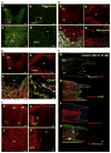
Experimental design and lesion extent. A. For Western blots, tissue blocks included 10 mm centered on the cervical or lumbar enlargement and 6 mm centered on the lesion epicenter (above diagram). For immunohistochemistry (IHC), spinal cords from PFA perfused rats were blocked with 10 mm centered on the cervical or lumbar enlargements or 20 mm centered on the epicenter. The epicenter IHC block was then severed at the lesion center and longitudinal or transverse cryostat sections were taken from the rostral and caudal ends. B. Eriochrome cyanine/cresyl violet (EC/CV) stained transverse sections from naïve thoracic spinal cord and the lesion epicenter of specimens obtained at 3, 7, 14, and 28 dpi, respectively. C, D. Midsagittal sections illustrating the extent of the spinal contusion site at 3 (C) and 28 dpi (D). In each pair, the top section was stained with EC/CV, and the bottom section with anti-GFAP. Arrow doublets (C) point to rostral and caudal lesion expansion in the dorsal column region. *(D) in GFAP-free lesion core; black arrows at GFAP+lesion border. Scale B=200 μm; C,D=1.0 mm.
Fig. 2
Fig. 2
Changes in CSPG core protein expression at the site of a severe contusion injury. A. Western blots of aggrecan, neurocan, brevican and NG2 in normal and injured thoracic cord. B. Analysis of identified bands of aggrecan, neurocan, brevican and NG2 expression in control (CTRL) and injured rats (designated dpi or DPI). Aggrecan expression changed with time (one way ANOVA; p<0.001) and decreased by 3 dpi, and remained absent through the remainder of the study. Total neurocan expression levels were increased 4 fold at 7 dpi and remain 3–4 fold elevated throughout 28 dpi. NG2 expression was not changed in the epicenter block. Post-hoc Dunnett’s *p<0.05; **p<0.01; ***p<0.001 compared with control.
Fig. 3
Fig. 3
IHC images of CSPG core proteins around the lesion epicenter after contusion injury. A. Aggrecan is expressed throughout the gray matter (dorsal horn, dh) and ventral horn (vh) in naïve spinal cord (a). The boxed area in (a) is enlarged in (c). Aggrecan is depleted throughout the epicenter (*) at 14 days after contusion injury (b). The boxed area in (b) is enlarged in (d) (ventrolateral white matter, vlwm). Scale a,b=200 μm; c,d=50 μm. B. (a) Neurocan (red) is expressed more highly in gray matter (dh, vh) than in white matter in naïve spinal cord. (b) Following injury, neurocan is upregulated throughout spared ventrolateral white matter regions (vlwm; 7 dpi). (c) Longitudinal section through the lesion border of another specimen at 7 dpi. The lesion (*) is at the top. (d) Scanning confocal micrograph from section at the lesion border at 7 dpi. Neurocan (red) is associated with hypertrophied astrocytes expressing high levels of GFAP (green). Scale a,b=200 μm, c=50 μm, d=10 μm. C. (a) Uninjured spinal cord. Brevican immunoreactivity is distributed throughout gray and white matter regions. (b) After injury, brevican staining intensity is increased throughout spared white and gray matter regions surrounding the lesion site (*) (14 dpi). (c) Enlargement of ventral horn from (a). (d) Enlargement of boxed area in (b) of ventrolateral white matter (vlwm) border and lesion epicenter (*) at 14 dpi. Scale a, b=200 μm, c=20 μm, d=50 μm. D. (a) NG2+ cells are distributed throughout gray and white matter regions in uninjured spinal cord. By 7 dpi (b), NG2 immunoreactivity is observed in hypertrophic NG2+ cells and in extracellular fibrillary profiles and other cell types. At 14 dpi, NG2+ cells are found in high density at the lesion border and extend far beyond the neurocan positive border (c), and into the center of the lesion (d). Scale a,b=200 μm, c=50 μm, d=40 μm. E. Photomontages of confocal images from adjacent dual-IHC stained sections of a specimen obtained at 14 dpi. The lesion epicenter is to the right of the panel (*) and the lesion border is outlined with a white line. Note that aggrecan staining is lost proximal to the lesion site (a), while neurocan and brevican (b,c) accumulate at the lesion border. NG2+ reactivity extends throughout the lesion center (d). Scale=400 μm.
Fig. 4
Fig. 4
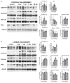
CSPG expression at cervical (A) and lumbar (B) levels. Western blots (left) and analysis of identified isoforms (right). Aggrecan and both isoforms of brevican expression were unchanged in cervical and lumbar spinal cord after contusion. Total and 250-kDa were changed in the cervical enlargement over time (one way ANOVA p<0.01 and p<0.0.02 respectively) with significant increase in total neurocan at 3 and 7 dpi (1.56 and 2.63 fold increases, respectively), and again, a large increased expression of the 250 kDa isoform (15.79 and 12,78 fold increases) at 3 and 7 dpi, respectively. Changes in the 150 kDa isoform of neurocan were modest, with a 1.58 fold increase compared with controls at 7 dpi. In the lumbar spinal cord, total and 250 kDa neurocan increased by 3 and 10–13 fold, respectively at 7 and 14 dpi; and both species remained elevated compared with controls for as long as 28 dpi. Significant increases in NG2 expression were observed at lumbar spinal cord levels (one way ANOVA; p<0.01) with a 2 fold increase (p >0.05) at 3 dpi and 3 fold increase (p<0.001) at 28 dpi. No differences in NG2 expression were observed at cervical spinal levels (p=0.076).
Fig. 5
Fig. 5
Regional expression of GFAP and neurocan in cervical and lumbar spinal cord. A. Distribution of GFAP+proportional area in white matter (top) and gray matter (bottom) regions. Top left, drawing of spinal cord enlargement with white matter regions of analysis; lateral dorsal columns (DC), medial dorsal columns (MDC), dorsal corticospinal tract (DCST), lateral white matter (LWM), and ventral white matter (VWM). Bottom left, gray matter regions; dorsal horn (DH), intermediate gray (IG), and ventral horn (VH). Graphs illustrate the proportional area measures across regions and time post-injury for cervical (center column) and lumbar (right column) enlargements. Two-way ANOVAs revealed a significant effect of region (p<0.001) and interaction (p<0.05) in both white and gray matter. Corrected post-hoc analyses showed increases in cervical MDC at 14 and 28 dpi and lumbar IG at 14 dpi (*p<0.05). B. Neurocan staining intensity in selected regions, including the medial dorsal columns (mdc), dorsal corticospinal tract (dcst), intermediate gray (ig), ventral gray matter (vgm) and lateral white matter (lwm). Two-way ANOVA identified effects of dpi and interaction in cervical and lumbar cord, with an increase in neurocan in cervical mdc at 14 and 28 dpi. In lumbar spinal cord, there was a significant increase in vgm at 28 dpi. Scale bar=200 μm.
Fig. 6
Fig. 6
Distribution of GFAP and neurocan in cervical dorsal columns and lumbar ventral gray matter. A. Dual staining images of GFAP, neurocan, and merged confocal photomicrographs illustrate chronic increase in neurocan immunohistochemical staining intensity in the medial dorsal columns of the cervical spinal cord. B. Increased neurocan staining is seen throughout the parenchyma of the lumbar spinal cord ventral gray matter. Scale bar=50 μm.

References

    1. Afshari FT, Kwok JC, White L, Fawcett JW. Schwann cell migration is integrin-dependent and inhibited by astrocyte-produced aggrecan. Glia. 2010;58(7):857–869. - PubMed
    1. Ankeny DP, McTigue DM, Jakeman LB. Bone marrow transplants provide tissue protection and directional guidance for axons after contusive spinal cord injury in rats. Exp Neurol. 2004;190 (1):17–31. - PubMed
    1. Asher RA, Morgenstern DA, Fidler PS, Adcock KH, Oohira A, Braistead JE, Levine JM, Margolis RU, Rogers JH, Fawcett JW. Neurocan is upregulated in injured brain and in cytokine-treated astrocytes. J Neurosci. 2000;20:2427–2438. - PMC - PubMed
    1. Barritt AW, Davies M, Marchand F, Hartley R, Grist J, Yip P, McMahon SB, Bradbury EJ. Chondroitinase ABC promotes sprouting of intact and injured spinal systems after spinal cord injury. J Neurosci. 2006;26:10856–10867. - PMC - PubMed
    1. Bekku Y, Rauch U, Ninomiya Y, Oohashi T. Brevican distinctively assembles extracellular components at the large diameter nodes of Ranvier in the CNS. J Neurochem. 2009;108:1266–1276. - PubMed

Publication types

Substances