. 2011;33(3-4):241-50.
doi: 10.1159/000331646. Epub 2011 Sep 27.

A novel approach to the study of hypoxia-ischemia-induced clinical and subclinical seizures in the neonatal rat

Affiliations

A novel approach to the study of hypoxia-ischemia-induced clinical and subclinical seizures in the neonatal rat

M Cuaycong et al. Dev Neurosci. 2011.

Abstract

Perinatal hypoxic-ischemic encephalopathy (HIE) is a major cause of acute mortality and chronic neurologic morbidity in infants and children. HIE is the most common cause of neonatal seizures, and seizure activity in neonates can be clinical, with both EEG and behavioral symptoms, subclinical with only EEG activity, or just behavioral. The accurate detection of these different seizure manifestations and the extent to which they differ in their effects on the neonatal brain continues to be a concern in neonatal medicine. Most experimental studies of the interaction between hypoxia-ischemia (HI) and seizures have utilized a chemical induction of seizures, which may be less clinically relevant. Here, we expanded our model of unilateral cerebral HI in the immature rat to include video EEG and electromyographic recording before, during and after HI in term-equivalent postnatal-day-12 rats. We observed that immature rats display both clinical and subclinical seizures during the period of HI, and that the total number of seizures and time to first seizure correlate with the extent of tissue damage. We also tested the feasibility of developing an automated seizure detection algorithm for the unbiased detection and characterization of the different types of seizure activity observed in this model.


Figures

Fig. 1
Fig. 1
A 30-second interval of EEG recording demonstrating a clinical seizure (EEG 2) defined as a recording with a rhythmic or repetitive tracing with an amplitude that increases to more than 3 times that of the baseline and lasting 3 s or longer. The top EEG trace (EEG 1) measures the potential between the left posterior and right anterior cortices. The middle EEG trace (EEG 2) measures the bilateral difference in potential in the parietal cortex. The bottom trace is the EMG recording from the upper extremities. Events such as this are compared with video to categorize them as clinical or subclinical seizures. Inset Video signal recorded in synchrony with EEG/EMG.
Fig. 2
Fig. 2
Time to first seizure was different between the 75-min group and the 90-min group. a Clinical. b Subclinical.
Fig. 3
Fig. 3
Brain damage as a function of duration of HI. Box-and-whisker plots demonstrating the range of damage, expressed as the infarct area, the percentage of the ipsilateral hemisphere relative to the contralateral hemisphere. The shorter duration, 75 min, resulted in a greater range of damage, whereas the more severe 90-min insult resulted in a greater number of severely damaged animals. The 2 animals with no damage were determined to be statistical outliers (more than 2 SD from the mean) and were not included in the analysis.
Fig. 4
Fig. 4
Hypoxic-ischemic brain damage as a function of numbers of seizures. Pearson correlation coefficients for these analyses are presented in table 3. The 2 animals with no damage in the 90-min group are more than 2 SD from the mean and were not included in the statistical analysis. a, b 90 min of HI. c, d 75 min of HI.
Fig. 5
Fig. 5
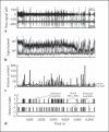
Sample seizure detection analysis for a P12 rat pup subjected to 90 min of HI. a Raw EEG and EMG signals during HI. EEG and EMG traces for the duration of hypoxia, with bursts of activity seen during possible seizures. b Mean signal power in a 1-second moving window. The signals are high-pass filtered at 5 Hz to reduce motion artifacts while amplifying high-frequency epileptiform spikes, and the mean square value in a moving 1-second window is computed. c Background correction and estimation of seizure content. The output is corrected for gross variations in the signal background, and a combination of rank filters (max-min operations) is applied to estimate the ‘seizure content’ of the signal in terms of the ratio of epileptiform activity normalized to background power. d Thresholding indicates periods of seizure activity. A threshold is applied to the EEG and EMG outputs in c to identify periods of time corresponding to candidates for subclinical seizures (EEG only), behavioral ‘seizures’ (EMG only) and clinical seizures (activity on both EEG and EMG). These candidates are verified visually by a human expert, and the performance of the algorithm is assessed using standard receiver operating characteristic analysis.

References

    1. Vannucci RC. Hypoxic-ischemic encephalopathy. Am J Perinatol. 2000;17:113–120. - PubMed
    1. Volpe JJ. Perinatal brain injury: from pathogenesis to neuroprotection. Ment Retard Dev Disabil Res Rev. 2001;7:56–64. - PubMed
    1. Cowan LD. The epidemiology of the epilepsies in children. Ment Retard Dev Disabil Res Rev. 2002;8:171–181. - PubMed
    1. Eicher DJ, Wagner CL, Katikaneni LP, Hulsey TC, Bass WT, Kaufman DA, Horgan MJ, Languani S., Bhatia JJ, Givelichian LM, Sankaran K., Yager JY. Moderate hypothermia in neonatal encephalopathy: safety outcomes. Pediatr Neurol. 2005;32:18–24. - PubMed
    1. Gluckman PD, Wyatt JS, Azzopardi D., Ballard R., Edwards AD, Ferriero DM, Polin RA, Robertson CM, Thoresen M., Whitelaw A., Gunn AJ. Selective head cooling with mild systemic hypothermia after neonatal encephalopathy: multicentre randomised trial. Lancet. 2005;365:663–670. - PubMed