

Early treatment of Class III malocclusion: 10-year clinical follow-up
- PMID: 21952927
- PMCID: PMC4223798
- DOI: 10.1590/s1678-77572011000400022
Early treatment of Class III malocclusion: 10-year clinical follow-up
Abstract
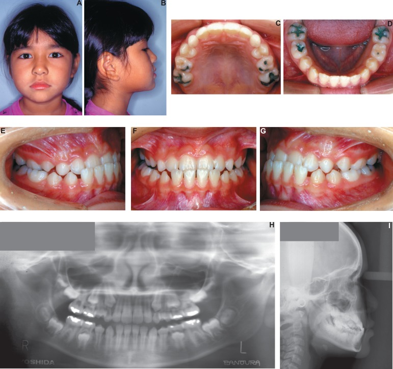
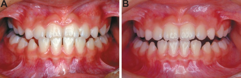
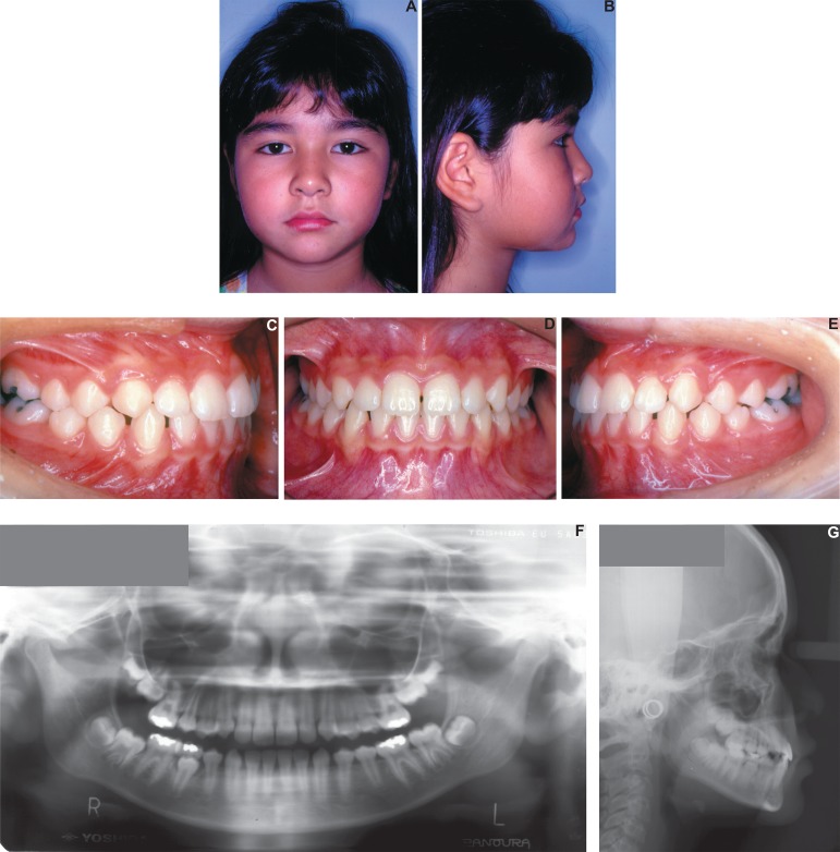
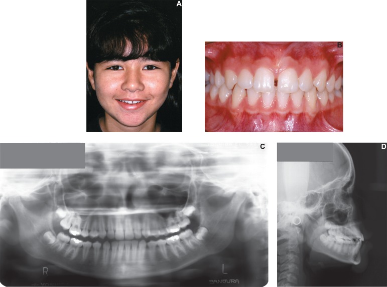
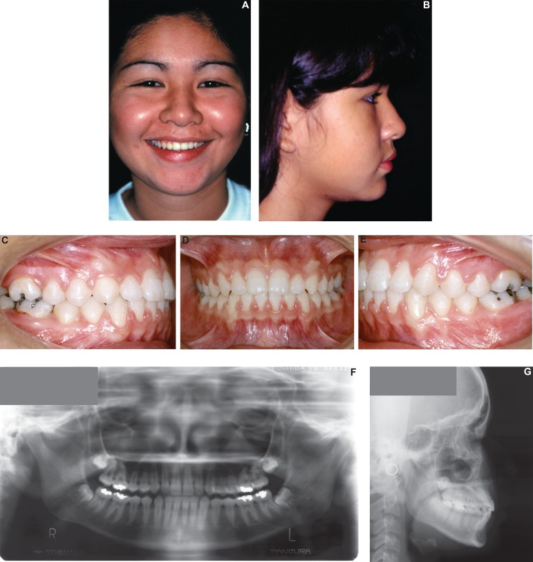
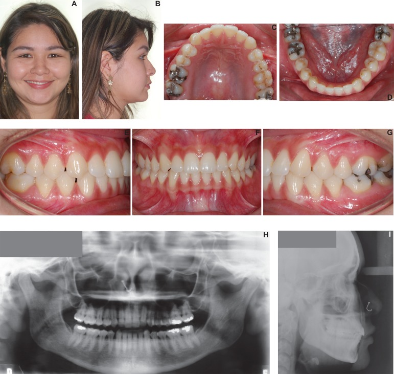
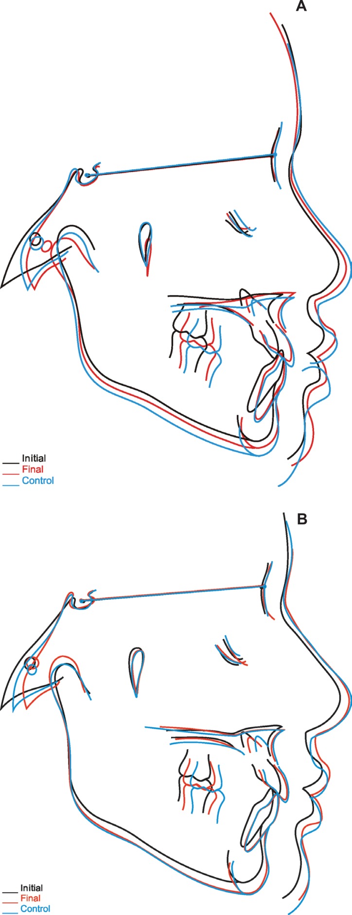
Angle Class III malocclusion has been a challenge for researchers concerning diagnosis, prognosis and treatment. It has a prevalence of 5% in the Brazilian population, and may have a genetic or environmental etiology. This malocclusion can be classified as dentoalveolar, skeletal or functional, which will determine the prognosis. Considering these topics, the aim of this study was to describe and discuss a clinical case with functional Class III malocclusion treated by a two-stage approach (interceptive and corrective), with a long-term follow-up. In this case, the patient was treated with a chincup and an Eschler arch, used simultaneously during 14 months, followed by corrective orthodontics. It should be noticed that, in this case, initial diagnosis at the centric relation allowed visualizing the anterior teeth in an edge-to-edge relationship, thereby favoring the prognosis. After completion of the treatment, the patient was followed for a 10-year period, and stability was observed. The clinical treatment results showed that it is possible to achieve favorable outcomes with early management in functional Class III malocclusion patients.
Figures

References
-
- Arun T, Nalbantgil D, Sayinsu K. Orthodontic treatment protocol of Ehlers-Danlos syndrome type VI. Angle Orthod. 2006;76(1):177–183. - PubMed
-
- Chung JC. Redirecting the growth pattern with rapid maxillary expander and chin cup treatment: changing breathing pattern from oral to nasal. World J Orthod. 2006;7(3):236–253. - PubMed
-
- Cozzani G. Extraoral traction and class III treatment. Am J Orthod. 1981;80(6):638–650. - PubMed
-
- Godt A, Zeyher C, Schatz-Maier D, Göz G. Early treatment to correct Class III relations with or without face masks. Angle Orthod. 2008;78(1):44–49. - PubMed
-
- Graber LW. Chin cup therapy for mandibular prognathism. Am J Orthod. 1977;72(1):23–41. - PubMed
Publication types
MeSH terms
LinkOut - more resources
Full Text Sources
