. 2011 Jun;22(6):997-1013.
doi: 10.1007/s13361-011-0117-9. Epub 2011 Apr 14.

Electron transfer dissociation of milk oligosaccharides

Affiliations

Electron transfer dissociation of milk oligosaccharides

Liang Han et al. J Am Soc Mass Spectrom. 2011 Jun.

Abstract

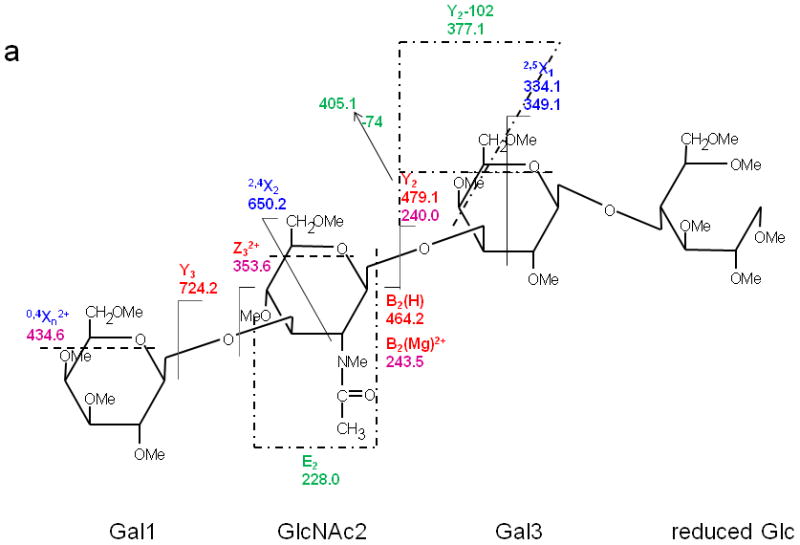
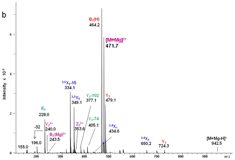
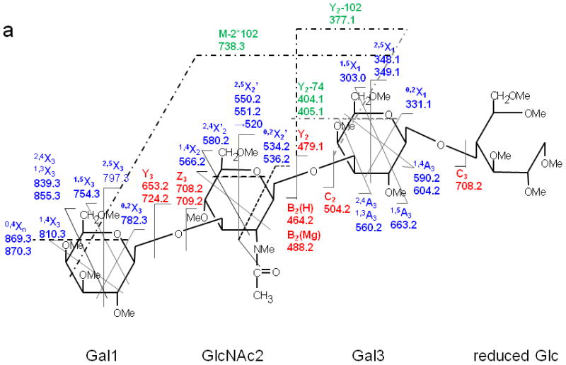
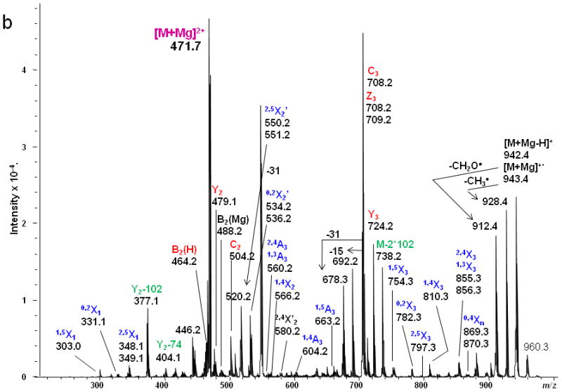
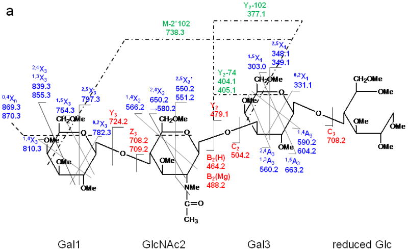
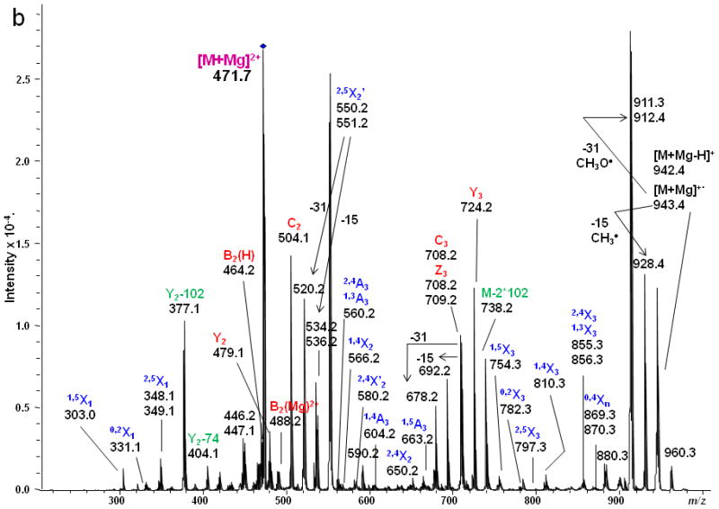
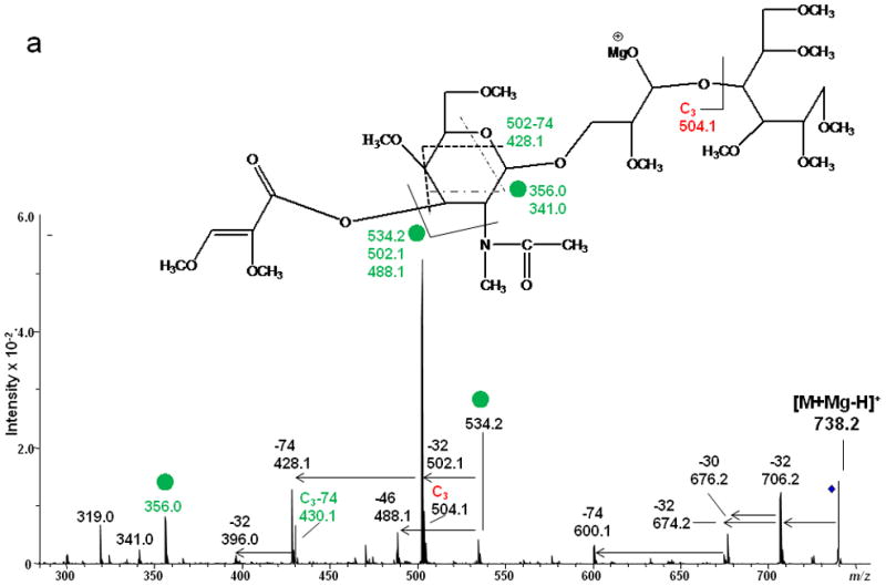
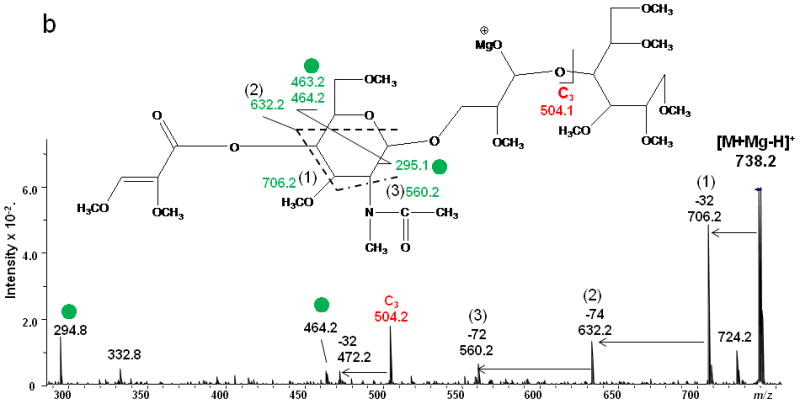
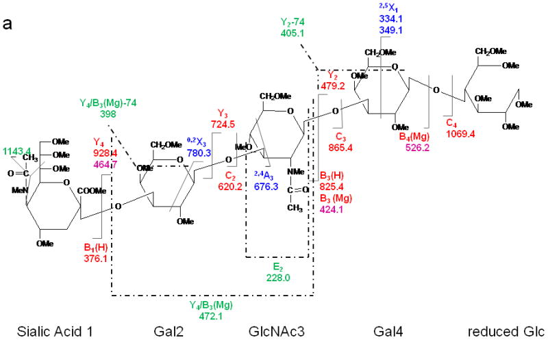
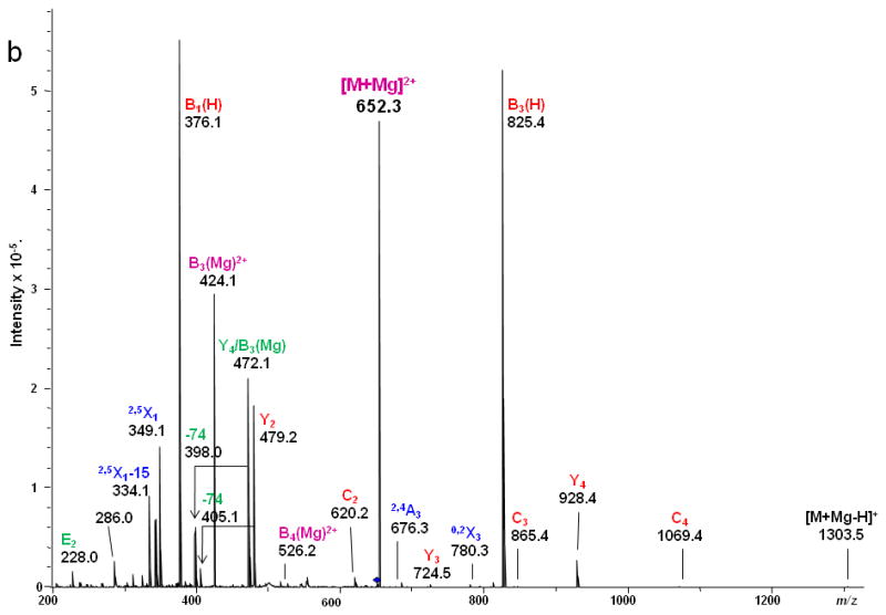
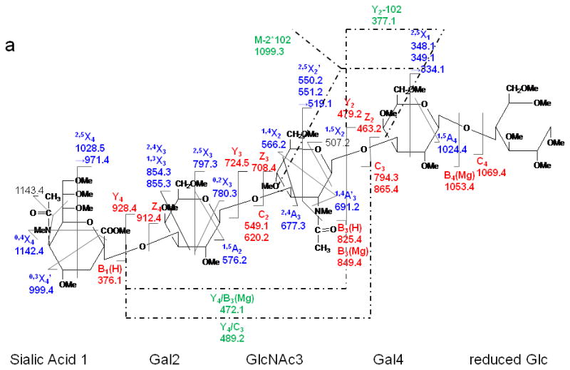
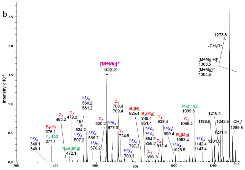
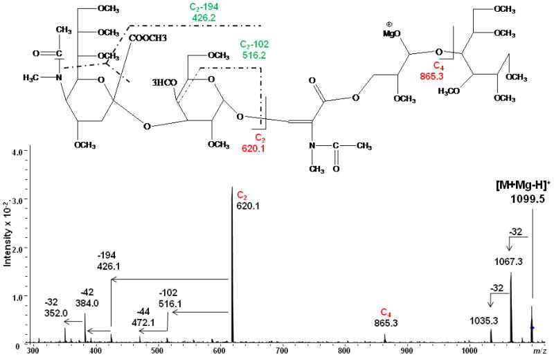
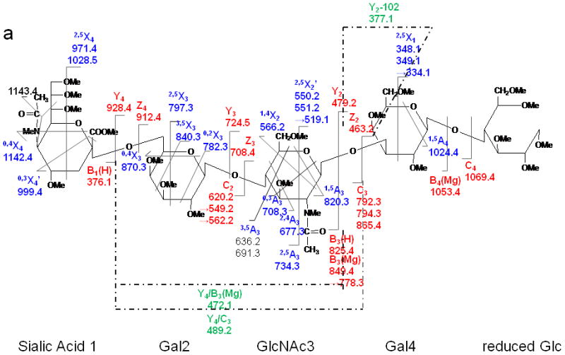
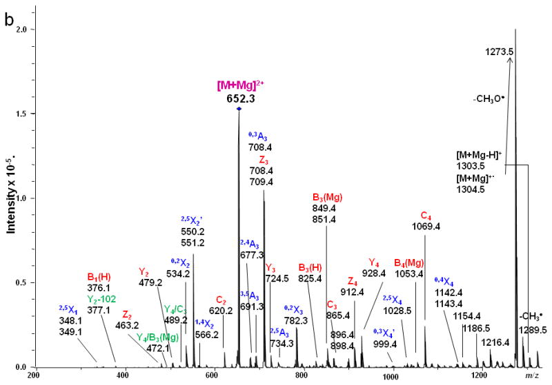
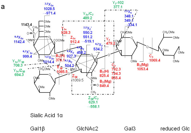
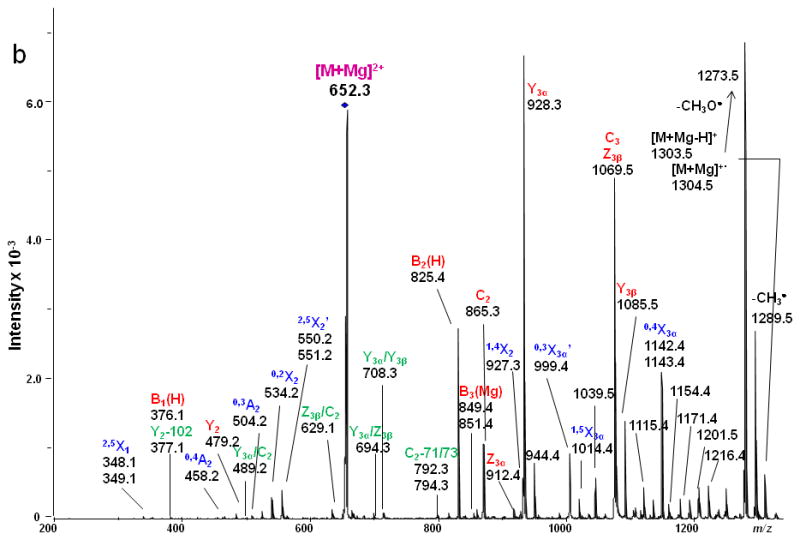
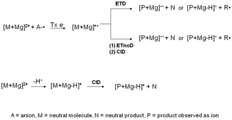
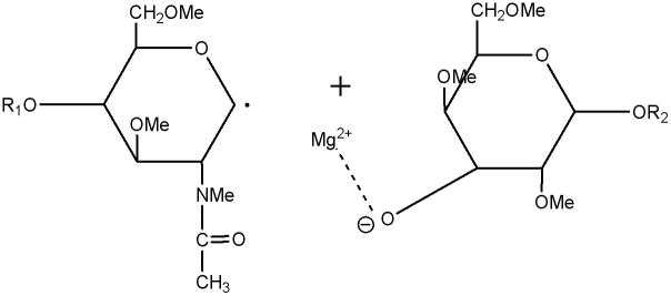
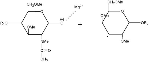
For structural identification of glycans, the classic collision-induced dissociation (CID) spectra are dominated by product ions that derived from glycosidic cleavages, which provide only sequence information. The peaks from cross-ring fragmentation are often absent or have very low abundances in such spectra. Electron transfer dissociation (ETD) is being applied to structural identification of carbohydrates for the first time, and results in some new and detailed information for glycan structural studies. A series of linear milk sugars was analyzed by a variety of fragmentation techniques such as MS/MS by CID and ETD, and MS(3) by sequential CID/CID, CID/ETD, and ETD/CID. In CID spectra, the detected peaks were mainly generated via glycosidic cleavages. By comparison, ETD generated various types of abundant cross-ring cleavage ions. These complementary cross-ring cleavages clarified the different linkage types and branching patterns of the representative milk sugar samples. The utilization of different MS(3) techniques made it possible to verify initial assignments and to detect the presence of multiple components in isobaric peaks. Fragment ion structures and pathways could be proposed to facilitate the interpretation of carbohydrate ETD spectra, and the main mechanisms were investigated. ETD should contribute substantially to confident structural analysis of a wide variety of oligosaccharides.


Figures

Figure 1
Figure 1
(a) CID MS/MS fragmentation of reduced and permethylated LNT [M + Mg]2+ m/z 471.7. (b) CID MS/MS spectrum. In this figure and all other figures, the ions that result from glycosidic cleavages have been labeled in red; those that originate from cross-ring cleavages have been labeled in blue, and those from internal cleavages, in green. The peaks labeled in purple represent doubly-charged ions.
Figure 1
Figure 1
(a) CID MS/MS fragmentation of reduced and permethylated LNT [M + Mg]2+ m/z 471.7. (b) CID MS/MS spectrum. In this figure and all other figures, the ions that result from glycosidic cleavages have been labeled in red; those that originate from cross-ring cleavages have been labeled in blue, and those from internal cleavages, in green. The peaks labeled in purple represent doubly-charged ions.
Figure 2
Figure 2
(a) ETD MS/MS fragmentation of reduced and permethylated LNT [M + Mg]2+ m/z 471.7. (b) ETD MS/MS spectrum.
Figure 2
Figure 2
(a) ETD MS/MS fragmentation of reduced and permethylated LNT [M + Mg]2+ m/z 471.7. (b) ETD MS/MS spectrum.
Figure 3
Figure 3
(a) ETD MS/MS fragmentation of reduced and permethylated LNnT [M + Mg]2+ m/z 471.7. (b) ETD MS/MS spectrum.
Figure 3
Figure 3
(a) ETD MS/MS fragmentation of reduced and permethylated LNnT [M + Mg]2+ m/z 471.7. (b) ETD MS/MS spectrum.
Figure 4
Figure 4
ETD/CID MS3 fragmentation and CID MS3 spectra of [M + Mg - H]+ m/z 738.2 generated via ETD of [M + Mg]2+ of (a) reduced and permethylated LNT and (b) reduced and permethylated LNnT.
Figure 4
Figure 4
ETD/CID MS3 fragmentation and CID MS3 spectra of [M + Mg - H]+ m/z 738.2 generated via ETD of [M + Mg]2+ of (a) reduced and permethylated LNT and (b) reduced and permethylated LNnT.
Figure 5
Figure 5
CID MS/MS (a) fragmentation and (b) spectrum of reduced and permethylated LSTa [M + Mg]2+ m/z 652.3.
Figure 5
Figure 5
CID MS/MS (a) fragmentation and (b) spectrum of reduced and permethylated LSTa [M + Mg]2+ m/z 652.3.
Figure 6
Figure 6
ETD MS/MS (a) fragmentation and (b) spectrum of reduced and permethylated LSTa [M + Mg]2+ m/z 652.3.
Figure 6
Figure 6
ETD MS/MS (a) fragmentation and (b) spectrum of reduced and permethylated LSTa [M + Mg]2+ m/z 652.3.
Figure 7
Figure 7
ETD/CID MS3 fragmentation and CID MS3 spectrum of the product ion [M + Mg - H]+ m/z 1099.5 that is generated via loss of H+ and two eliminations of CH3OCH=CHCH2OCH3 (2x10² Da) in the ETD MS/MS spectrum of the [M + Mg]2+ of reduced and permethylated LSTa
Figure 8
Figure 8
ETD MS/MS (a) fragmentation and (b) spectrum of reduced and permethylated LSTc [M + Mg]2+ m/z 652.3. Fragments between m/z 1142.2 and 1216.4 are attributed to cleavages within the sialic acid residue.
Figure 8
Figure 8
ETD MS/MS (a) fragmentation and (b) spectrum of reduced and permethylated LSTc [M + Mg]2+ m/z 652.3. Fragments between m/z 1142.2 and 1216.4 are attributed to cleavages within the sialic acid residue.
Figure 9
Figure 9
ETD MS/MS (a) fragmentation and (b) spectrum of reduced and permethylated LSTb [M + Mg]2+ m/z 652.3. Fragments between m/z 1115.4 and 1216.4 are attributed to cleavages within the sialic acid residue.
Figure 9
Figure 9
ETD MS/MS (a) fragmentation and (b) spectrum of reduced and permethylated LSTb [M + Mg]2+ m/z 652.3. Fragments between m/z 1115.4 and 1216.4 are attributed to cleavages within the sialic acid residue.
Scheme 1
Scheme 1
Scheme 2
Scheme 2
Y-type ETD ion and neutral radical
Scheme 3
Scheme 3
C-type ETD product ion and neutral radical
Scheme 4
Scheme 4
(a) Singly-charged even-electron Z-type ions and (b) singly-charged odd-electron Z-type ions
Scheme 5
Scheme 5
Suggested structures of (a) [Bn(H)]+ and (b) [Bn(Mg)+2H]+- ions.
Scheme 6
Scheme 6
Suggested structures of 2,5Xn ETD product ions and neutral loss
Scheme 7
Scheme 7
Suggested route for formation of 2,5Xn' ETD product ions
Scheme 8
Scheme 8
Suggested structures of 0,3An ETD product ion and neutral radical

References

    1. Ohtsubo K, Marth JD. Glycosylation in cellular mechanisms of health and disease. Cell. 2006;126:855–867. - PubMed
    1. Varki A, Cummings RD, Esko JD, Freeze HH, Hart GW, Marth J, editors. Essentials of Glycobiology. Cold Spring Harbor Laboratory Press; Cold Spring Harbor, New York: 1999. - PubMed
    1. Mutenda KE, Matthiesen R. Analysis of carbohydrates by mass spectrometry. In: Matthiesen R, editor. Mass Spectrometry Data Analysis in Proteomics. Humana Press; Totowa, New Jersey: 2006. p. 289.
    1. Xie B, Costello CE. Carbohydrate structure determination by mass spectrometry. In: Cowman MK, Garg HG, Hales CA, editors. Carbohydrate Chemistry, Biology and Medical Applications. Elsevier, Ltd; New York: 2008. p. 29.
    1. Ashline D, Singh S, Hanneman A, Reinhold V. Congruent strategies for carbohydrate sequencing. 1. Mining structural details by MSn. Anal Chem. 2005;77:6250–6262. - PMC - PubMed

Publication types