Review
. 2012 Jan;226(2):316-25.
doi: 10.1002/path.2999. Epub 2011 Nov 14.

Nuclear lamins and laminopathies

Affiliations
Review

Nuclear lamins and laminopathies

Howard J Worman. J Pathol. 2012 Jan.

Abstract

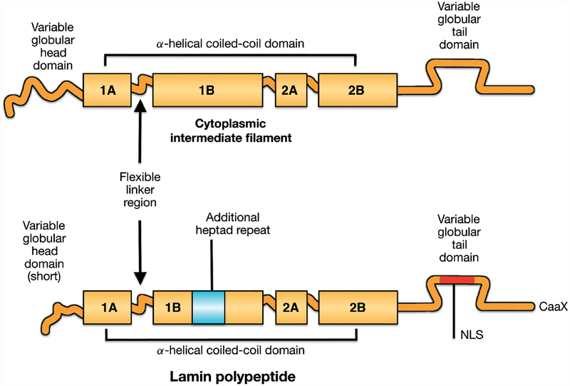
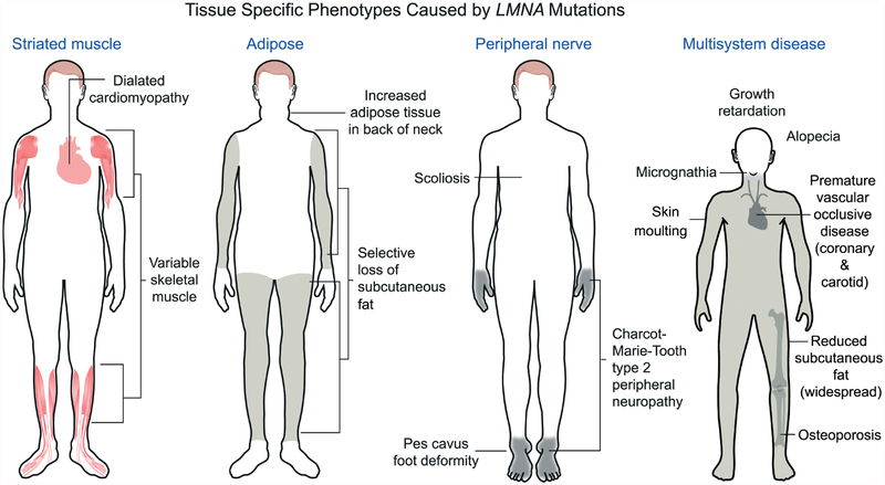
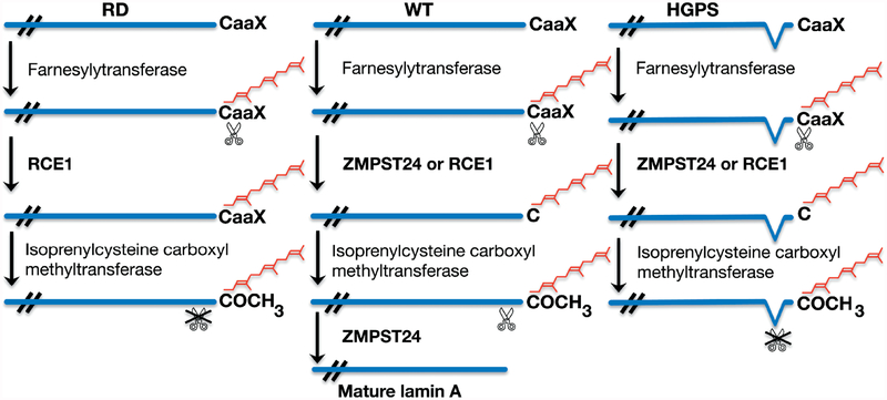
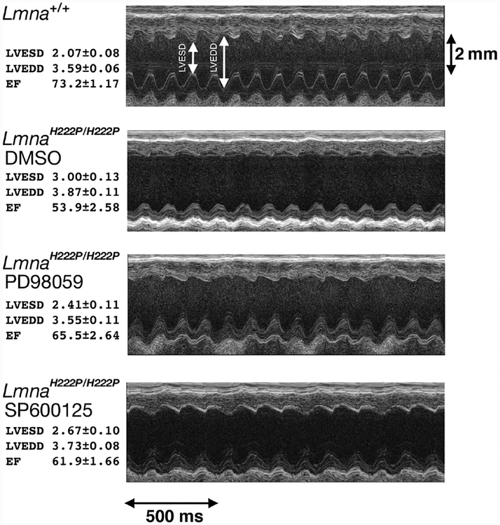
Nuclear lamins are intermediate filament proteins that polymerize to form the nuclear lamina on the inner aspect of the inner nuclear membrane. Long known to be essential for maintaining nuclear structure and disassembling/reassembling during mitosis in metazoans, research over the past dozen years has shown that mutations in genes encoding nuclear lamins, particularly LMNA encoding the A-type lamins, cause a broad range of diverse diseases, often referred to as laminopathies. Lamins are expressed in all mammalian somatic cells but mutations in their genes lead to relatively tissue-selective disease phenotypes in most cases. While mutations causing laminopathies have been shown to produce abnormalities in nuclear morphology, how these disease-causing mutations or resultant alterations in nuclear structure lead to pathology is only starting to be understood. Despite the incomplete understanding of pathogenic mechanisms underlying the laminopathies, basic research in cellular and small animal models has produced promising leads for treatments of these rare diseases.


Conflict of interest statement

Conflict of interest: The author is an inventor on a pending PCT patent application on methods for treating and/or preventing cardiomyopathies by ERK and JNK inhibition, filed by the Trustees of Columbia University in the City of New York.

Figures

Figure 1.
Figure 1.
Schematic diagrams showing vertebrate intermediate filament protein structures and differences between cytoplasmic intermediate filament proteins and nuclear lamins. All intermediate filament proteins, including lamins, have a conserved domain structure consisting a central α-helical coiled-coil rod domain consisting of four coiled coils (1A, 1B, 2A, 2B), based on heptad repeats, interrupted by flexible linker domains and a variable globular tail domain. Compared to vertebrate cytoplasmic intermediate filament proteins, lamins contain six additional heptad repeats (42 amino acids) in coil 1B, a nuclear localization signal (NLS) near an immunoglobulin-like fold domain in the carboxyl-terminal tail and, in most lamins, a CaaX motif at the carboxyl-terminus. Reproduced with permission from Hutchison CJ, Worman HJ. A-type lamins: guardians of the soma? Nat Cell Biol 2004; 6: 1062–1067 [1].
Figure 2.
Figure 2.
Different LMNA mutations cause diseases that affect striated muscle, adipose, peripheral nerve or multiple systems with features of accelerated ageing. Most LMNA mutations are autosomal-dominant and cause dilated cardiomyopathy with variable skeletal muscle involvement. This includes the classical Emery–Dreifuss muscular dystrophy phenotype, as shown in the diagram, with scapulohumeral–peroneal distribution of skeletal muscle involvement, concurrent tendon contractures and dilated cardiomyopathy. Autosomal-dominant missense mutations in LMNA, the large majority of which cause a change in the surface charge of the immunoglobulin-like fold of lamin A and lamin C, cause Dunnigan-type familial partial lipodystrophy, with selective loss of subcutaneous fat from the extremities, fat accumulation in the neck and face and insulin resistance and diabetes mellitus. An autosomal recessive LMNA mutation that leads to an arginine-to-cysteine amino acid substitution at residue 298 causes a Charcot–Marie–Tooth type 2 peripheral neuropathy, characterized clinically by a stocking–glove sensory neuropathy, resultant pes cavus foot deformity and other variable features, such as scoliosis. The sporadic cytosine to thymine transversion in codon 608 of exon 11 of LMNA causes Hutchinson–Gilford progeria syndrome, which has features of accelerated ageing, such as sclerotic skin, joint contractures, micrognathia, alopecia, fingertip tufting, distal-joint abnormalities, growth impairment and vascular abnormalities, generally leading to death during the second decade due to myocardial infarction or stroke. Other LMNA mutations can also cause progeroid syndromes with similar features; a recessive LMNA mutation causing an arginine-to-histidine amino acid substitution at residue 527 in the immunoglobulin-like fold causes mandibuloacral dysplasia, a disorder with a combination of progeroid features and partial lipodystrophy. Reproduced with permission from: Dauer WT, Worman HJ. The nuclear envelope as a signalling node in development and disease. Dev Cell 2009; 17: 626–638 [65].
Figure 3.
Figure 3.
Processing of prelamin A in wild-type (WT) cells occurs in a series of sequential enzymatic reactions that lead to farnesylation, endoproteolytic cleavage of–aaX, carboxymethylation and a second endoproteolytic cleavage catalysed by ZMSTE24. In restrictive dermopathy (RD), there is no ZMPSTE24 activity, which results in accumulation of farnesylated, carboxymethylated prelamin A. In Hutchinson–Gilford progeria syndrome (HGPS), the second site for cleavage catalysed by ZMPSTE24 is deleted, which leads to accumulation of progerin, a truncated variant of farnesylated, carboxymethylated prelamin A. Reproduced with permission from: Worman HJ, Östlund C, Wang Y. Diseases of the nuclear envelope. Cold Spring Harb Perspect Biol 2010; 2: a000760 [66].
Figure 4.
Figure 4.
Representative transthoracic M-mode echocardiograms taken from wild-type mice (Lmna+/+), Lmna mutant mice that develop cardiomyopathy receiving placebo (LmnaH222P/H222P DMSO), Lmna mutant mice treated with an inhibitor of extracellular signal-regulated kinase signalling (LmnaH222P/H222P PD98059) and Lmna mutant mice treated with an inhibitor of c-Jun N-terminal kinase signalling (LmnaH222P/H222P SP600125). Left ventricular end systolic diameter (LVESD) and left ventricular end diastolic diameter (LVEDD) are indicated in the top echocardiographic tracing. At left, means ± standard errors for LVESD, LVEDD and the cardiac ejection fraction (EF), a measure of cardiac contractility, are given for mice in each group. Both PD98059 and SP600125 significantly improve the ejection fraction compared to placebo. This figure is based on Figure 3 and data are used with permission from: Wu W, Muchir A, Shan J, et al. Mitogen-activated protein kinase inhibitors improve heart function and prevent fibrosis in cardiomyopathy caused by mutation in lamin A/C gene. Circulation 2011; 123: 53–61 [108].

References

    1. Melcer S, Gruenbaum Y, Krohne G. Invertebrate lamins. Exp Cell Res 2007; 313: 2157–2166. - PubMed
    1. Hutchison CJ, Worman HJ. A-type lamins: guardians of the soma? Nat Cell Biol 2004; 6: 1062–1067. - PubMed
    1. Frangioni J, Neel B Use of a general purpose mammalian expression vector for studying intracellular protein targeting: identification of critical residues in the nuclear lamin A/C nuclear localisation sequence. J Cell Sci 1993; 105: 481–488. - PubMed
    1. Dhe-Paganon S, Werner ED, Chi YI, et al. Structure of the globular tail of nuclear lamin. J Biol Chem 2002; 277: 17381–17384. - PubMed
    1. Krimm I, Östlund C, Gilquin B, et al. The Ig-like structure of the C-terminal domain of lamin A/C, mutated in muscular dystrophies, cardiomyopathy, and partial lipodystrophy. Structure 2002; 10: 811–823. - PubMed

Publication types