. 2012 Mar;71(3):424-31.
doi: 10.1136/ard.2011.154211. Epub 2011 Sep 27.

Histone deacetylase inhibitors suppress rheumatoid arthritis fibroblast-like synoviocyte and macrophage IL-6 production by accelerating mRNA decay

Affiliations
Free PMC article

Histone deacetylase inhibitors suppress rheumatoid arthritis fibroblast-like synoviocyte and macrophage IL-6 production by accelerating mRNA decay

Aleksander M Grabiec et al. Ann Rheum Dis. 2012 Mar.
Free PMC article

Abstract

Background: Histone deacetylase inhibitors (HDACi) display potent therapeutic efficacy in animal models of arthritis and suppress inflammatory cytokine production in rheumatoid arthritis (RA) synovial macrophages and tissue.

Objectives: To determine the molecular mechanisms contributing to the suppressive effects of HDACi on RA synovial cell activation, using interleukin 6 (IL-6) regulation as a model.

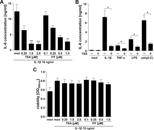
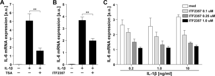
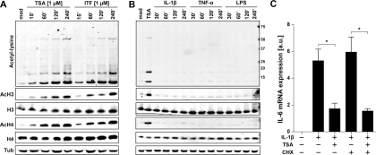
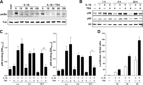
Methods: RA fibroblast-like synoviocytes (FLS) and healthy donor macrophages were treated with IL-1β, tumour necrosis factor (TNF)α, lipopolysaccharide or polyinosinic:polycytidylic acid (poly(I:C)) in the absence or presence of the HDACi trichostatin A (TSA) or ITF2357 (givinostat). IL-6 production and mRNA expression was measured by ELISA and quantitative PCR (qPCR), respectively. Protein acetylation and the activation of intracellular signalling pathways were assessed by immunoblotting. The DNA-binding activity of nuclear factor κB (NFκB) and activator protein 1 (AP-1) components was measured by ELISA-based assays.

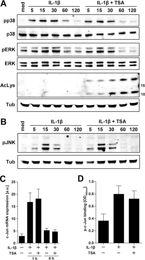
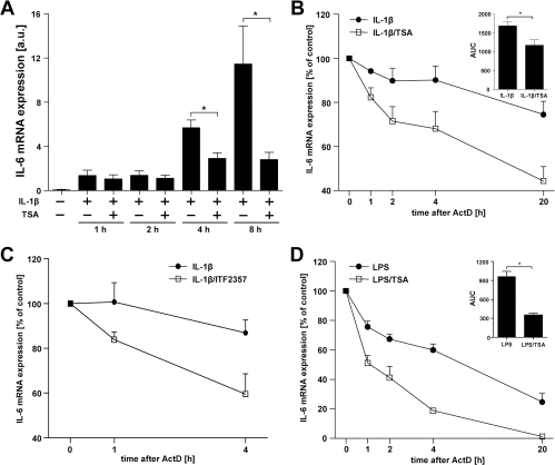
Results: HDACi (0.25-1.0 μM) suppressed RA FLS IL-6 production induced by IL-1β, TNFα and Toll-like receptor ligands. Phosphorylation of mitogen-activated protein kinases and inhibitor of κBα (IκBα) following IL-1β stimulation were unaffected by HDACi, as were AP-1 composition and binding activity, and c-Jun induction. TSA induced a significant reduction in nuclear retention of NFκB in FLS 24 h after IL-1β stimulation, but this did not reduce NFκB transcriptional activity or correlate temporally with reductions in IL-6 mRNA accumulation. HDACi significantly reduced the stability of IL-6 mRNA in FLS and macrophages.

Conclusions: Our study identifies a novel, shared molecular mechanism by which HDACi can disrupt inflammatory cytokine production in RA synovial cells, namely the promotion of mRNA decay, and suggests that targeting HDAC activity may be clinically useful in suppressing inflammation in RA.


Figures

Figure 1
Figure 1
Histone deacetylase inhibitors (HDACi) suppress interleukin 6 (IL-6) production by rheumatoid arthritis fibroblast-like synoviocytes (FLS) without affecting cell viability. A. FLS were stimulated with 10 ng/ml of IL-1β alone or in the presence of increasing concentrations of trichostatin A (TSA) or ITF2357 for 24 h (n=6–10). B. Alternatively, cells were left untreated or pre-exposed to 1 µM TSA followed by 24 h stimulation with IL-1β (10 ng/ml), tumour necrosis factor (TNF)α (10 ng/ml), lipopolysaccharide (1 µg/ml) or polyinosinic:polycytidylic acid (poly(I:C)) (10 µg/ml) (n=4). IL-6 levels in cell-free tissue culture supernatants were determined by ELISA. Results, presented as mean±SEM IL-6 concentration, were either subjected to the Kruskal–Wallis test followed by Dunns' multiple comparison analysis with cells not treated with HDACi used as reference controls (A), or to the Mann–Whitney U test (B). *p<0.05; **p<0.01; ***p<0.001. C. Viability of cells after 24 h stimulation with IL-1β (10 ng/ml) in the presence or absence of increasing doses of TSA or ITF2357 was assessed by measurement of 3-(4,5-dimethylthiazol-2-yl)-2,5-diphenyltetrazolium bromide (MTT) reduction (n=5). Values representing changes in MTT processing are shown as mean±SEM optical density at 590 nm.
Figure 2
Figure 2
Histone deacetylase inhibitors reduce interleukin 6 (IL-6) mRNA accumulation in rheumatoid arthritis fibroblast-like synoviocytes (FLS). FLS were left unstimulated, or were stimulated with IL-1β (10 ng/ml) with or without 1 µM trichostatin A (A), or with IL-1β (1 ng/ml) in the presence or absence of 0.25 µM ITF2357 (B) for 4 h, total RNA was extracted, reverse transcribed and IL-6 mRNA accumulation analysed by quantitative PCR (qPCR). Results are presented as mean±SEM IL-6 expression relative to 18S of five independent experiments. **p<0.01, Mann–Whitney U test. Alternatively (C), FLS (n=3) were stimulated as above with increasing concentrations of IL-1β (0.2–10 ng/ml) in the absence (med) or presence of increasing concentrations of ITF2357 (0.1–1.0 μM) and IL-6 mRNA expression assessed as in (A) and (B).
Figure 3
Figure 3
New protein synthesis is not required for fibroblast-like synoviocyte (FLS) interleukin 6 (IL-6) suppression by histone deacetylase inhibitors. A. FLS were left untreated (med) or were treated with trichostatin A (TSA) (1 µM) or ITF2357 (1 µM) for the indicated time (min). Protein extracts were prepared and analysed by immunoblotting for acetylated lysine (Ac-Lys), Ac-H3, H3, Ac-H4, H4 and tubulin (Tub) content. B. FLS were left unstimulated in medium (med) or were stimulated with IL-1β (10 ng/ml), tumour necrosis factor (TNF)α (10 ng/ml) or lipopolysaccharide (1 µg/ml), lysed and examined by immunoblotting with antibodies recognising Ac-Lys, Ac-H3, H3, Ac-H4, H4 and Tub. FLS treated with 1 µM TSA for 4 h were used as a positive control. Results are representative of three independent experiments. C. FLS were left untreated or were treated with IL-1β (10 ng/ml) with or without 1 µM TSA for 4 h. To block protein translation, cells were preincubated with 1 µg/ml cycloheximide (CHX) for 30 min prior to stimulation. IL-6 mRNA levels were assessed by quantitative PCR (qPCR) and presented as relative IL-6 expression ±SEM (n=4). *p<0.05, Mann–Whitney U test.
Figure 4
Figure 4
Histone deacetylase inhibitors (HDACi) fail to affect mitogen-activated protein kinase (MAPK) signalling and activator protein 1 (AP-1) activation in rheumatoid arthritis fibroblast-like synoviocytes (FLS). A. FLS were left unstimulated (med) or were stimulated with 10 ng/ml of interleukin 1β (IL-1β) for the indicated time (min) in the presence or absence of 1 µM trichostatin A (TSA), proteins were extracted and MAPK activation analysed by immunoblotting with antibodies specific for phospho(p)-p38, p38, p-extracellular signal regulated kinase (ERK), ERK and tubulin (Tub). Anti-acetylated lysine (Ac-Lys) antibody was used to confirm TSA HDACi activity in this experiment. B. FLS were subjected to the same treatment as in (A) and c-Jun N-terminal kinase (JNK) activation determined in cellular lysates by immunoblotting for p-JNK and Tub content. C. HDACi do not modulate c-Jun induction by an inflammatory stimulus. FLS were stimulated with medium alone (med) or with IL-1β (10 ng/ml) with or without 1 µM TSA for 1 h or 4 h, total RNA was reverse transcribed, and changes in c-Jun mRNA levels were determined by quantitative PCR (qPCR). Results of four independent experiments are shown as relative expression ±SEM. D. HDACi have no effect on c-Jun DNA binding activity. FLS were left unstimulated in medium (med) or were treated with IL-1β (10 ng/ml) for 4 h in the presence or absence of 1 µM TSA, nuclear fractions were extracted and levels of active p-c-Jun were determined using an ELISA-based DNA-binding assay. Data represent the mean±SEM optical density at 450 nm for five independent experiments.
Figure 5
Figure 5
Histone deacetylase inhibitors (HDACi) increase nuclear factor κB (NFκB) transcriptional activity early after stimulation, but inhibit NFκB nuclear retention at later time points. A. Fibroblast-like synoviocytes (FLS) were left untreated (med) or were treated with interleukin 1β (IL-1β) (10 ng/ml) for 5–120 min in the presence or absence of 1 µM trichostatin A (TSA), and total cell lysates examined by immunoblotting for phospho(p)- inhibitor of κBα (IκBα) and tubulin (Tub) content. Alternatively, FLS were cultured in the presence or absence of 1 µM TSA and then stimulated with medium alone or 10 ng/ml of IL-1β for 1 h, 4 h or 24 h. Nuclear extracts (n=4) were either separated by electrophoresis and nuclear accumulation of NFκB subunits analysed by immunoblotting with antibodies recognising p50, p65 and histone 3 (H3) (B), or were used for determination of p50 and p65 DNA-biding activities by an ELISA-based DNA biding assay (C). Values representing changes in p50 (left panel) and p65 (right panel) are shown as mean±SEM optical density at 450 nm. *p<0.05; **p<0.01, Mann–Whitney U test. D. FLS were cotransduced with a 4×NFκB-minimal promoter (MLP)-luciferase and control cytomegalovirus β-galactosidase (CMV-β-gal) adenoviral vectors. At 48 h after transduction, cells were serum-starved for 24 h and then left untreated or stimulated with IL-1β (10 ng/ml) with or without 1 µM TSA for 2 h, 4 h or 8 h. Cellular lysates were prepared and the luciferase (Luc) and β-gal enzymatic activities determined by luminometry and spectrophotometry, respectively. Data are presented as relative luciferase activity normalised to β-gal activity ±SD and a representative of two independent experiments is shown.
Figure 6
Figure 6
Histone deacetylase inhibitors (HDACi) accelerate interleukin 6 (IL-6) mRNA degradation in rheumatoid arthritis fibroblast-like synoviocytes (FLS) and healthy donor (HD) macrophages. A. FLS were left untreated (med) or were treated with IL-1β (10 ng/ml) with or without 1 µM trichostatin A (TSA) for the indicated time (h), total RNA was extracted, cDNA was synthesised and temporal changes in IL-6 mRNA accumulation were monitored by quantitative PCR (qPCR). Data represent the mean±SEM of five or six independent experiments. *p<0.05, Mann–Whitney U test. B. FLS were stimulated with IL-1β (10 ng/ml) in the presence or absence of 1 µM TSA (n=5) or (C) with IL-1β (1 ng/ml) in the presence or absence of 250 nM ITF2357 (n=3). Alternatively, (D) monocyte-derived HD macrophages (n=4) were stimulated with lipopolysaccharide (1 µg/ml). After 4 h of stimulation cells were washed and fresh medium containing 10 µg/ml of actinomycin D (ActD) was added. RNA was extracted at the indicated time points (h) from the beginning of ActD treatment and the rates of IL-6 mRNA degradation in the presence or absence of HDACi were examined by qPCR. Values for the 0 h time point were normalised to 100%, and remaining values were expressed as the mean±SEM percentage of IL-6 mRNA levels compared with controls. For (B) and (D) the areas under the curves (AUCs) obtained for cells treated with an inflammatory stimulus alone or in the presence of TSA were calculated and differences between AUC values were analysed by the Mann–Whitney U test (insets, mean AUC±SEM). *p<0.05.

Comment in

References

    1. Klareskog L, Catrina AI, Paget S. Rheumatoid arthritis. Lancet 2009;373:659–72 - PubMed
    1. Bartok B, Firestein GS. Fibroblast-like synoviocytes: key effector cells in rheumatoid arthritis. Immunol Rev 2010;233:233–55 - PMC - PubMed
    1. Müller-Ladner U, Ospelt C, Gay S, et al. Cells of the synovium in rheumatoid arthritis. Synovial fibroblasts. Arthritis Res Ther 2007;9:223. - PMC - PubMed
    1. Urnov FD, Wolffe AP. Chromatin remodeling and transcriptional activation: the cast (in order of appearance). Oncogene 2001;20:2991–3006 - PubMed
    1. de Ruijter AJ, van Gennip AH, Caron HN, et al. Histone deacetylases (HDACs): characterization of the classical HDAC family. Biochem J 2003;370:737–49 - PMC - PubMed

MeSH terms