. 2011 Nov 11;286(45):39282-9.
doi: 10.1074/jbc.M111.253807. Epub 2011 Sep 27.

The Fbw7 tumor suppressor regulates nuclear factor E2-related factor 1 transcription factor turnover through proteasome-mediated proteolysis

Affiliations

The Fbw7 tumor suppressor regulates nuclear factor E2-related factor 1 transcription factor turnover through proteasome-mediated proteolysis

Madhurima Biswas et al. J Biol Chem. .

Abstract

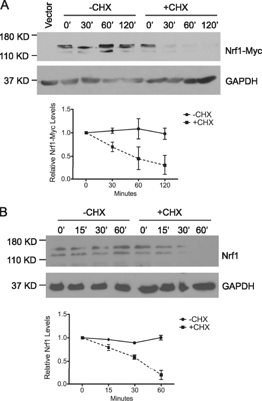
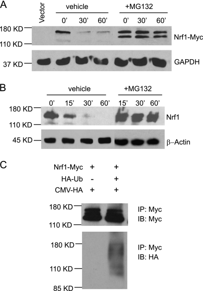
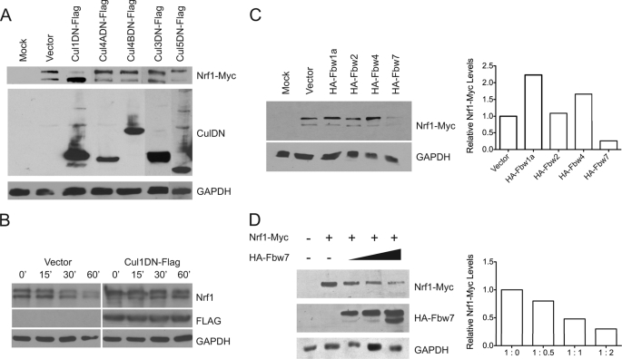
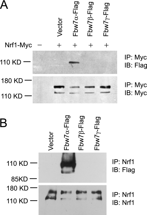
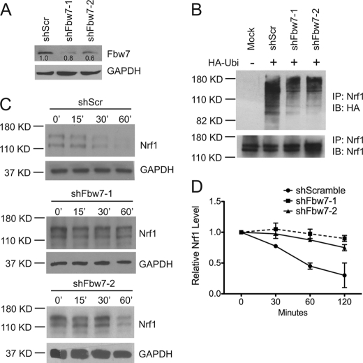
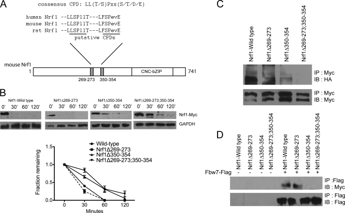
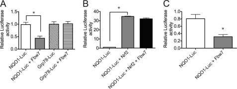
Nuclear factor E2-related factor 1 (Nrf1) is a basic leucine zipper transcription factor that plays important roles in cellular stress response and development. Currently, the mechanism regulating Nrf1 expression is poorly understood. We report here that Nrf1 is a short-lived protein that is targeted by F-box protein Fbw7, which is the substrate-specifying component of SCF (Skp1-Cul1-Fbox protein-Rbx1)-type ubiquitin ligase for degradation via the ubiquitin-proteasome pathway. We show that Fbw7 directly binds Nrf1 through a Cdc4 phosphodegron and that enforced expression of Fbw7 promotes the ubiquitination and degradation of Nrf1. Conversely, depletion of endogenous Fbw7 leads to decreased Nrf1 ubiquitination and accumulation of Nrf1 protein. Accordingly, expression of Fbw7 leads to down-regulation of antioxidant response element-driven gene activation, whereas disruption of Fbw7-mediated destabilization of Nrf1 leads to increased antioxidant response element-driven gene expression. Together, these data identify Fbw7 as a regulator of Nrf1 expression and reveal a novel function of Fbw7 in cellular stress response.


Figures

FIGURE 1.
FIGURE 1.
Nrf1 has a short half-life. A, HEK293 cells were transfected with vector or Myc-tagged Nrf1, followed by treatment with 50 μg/ml cycloheximide (CHX) for 0, 30, 60, and 120 min. Western blot analysis was done on the whole cell lysates using anti-Myc antibody. GAPDH was used as the loading control. The graph shows quantitative analysis of CHX chase data. Each point represents the mean (± S.E.) of the remaining protein. B, HEK293 cells were treated with 50 μg/ml CHX for 0, 30, 60, and 120 min. Western blot analysis was done on the whole cell lysates for detection of endogenous Nrf1 using anti-Nrf1 antibody. GAPDH was used as the loading control. The graph shows quantitative analysis of cycloheximide chase data. Each point represents the mean (± S.E.) of the remaining protein.
FIGURE 2.
FIGURE 2.
Nrf1 is ubiquitinated and stabilized by proteasomal inhibition. A, HEK293 cells were transfected with Nrf1-Myc, followed by treatment with vehicle (DMSO) or 10 μm of the proteasomal inhibitor, MG132, for 5 h followed by treatment with 50 μg/ml CHX. Cells were harvested at 0, 30, and 60 min, and Western blotting was done using an anti-Myc antibody. GAPDH was used as the loading control. B, HEK293 cells were treated vehicle (DMSO) or 10 μm of the proteasomal inhibitor, MG132, for 5 h followed by treatment with 50 μg/ml CHX. Cells were harvested at 0, 30, and 60 min, and Western blotting was done on endogenous Nrf1 using anti-Nrf1 antibody. GAPDH was used as the loading control. C, HEK293 cells were transfected with Nrf1-Myc, HA-Ub, and CMV-HA. Cell extracts were immunoprecipitated with an anti-Myc antibody and analyzed by immunoblotting with an anti-ubiquitin antibody. For input control, filter was blotted with anti-Myc antibody.
FIGURE 3.
FIGURE 3.
Nrf1 is destabilized by SCFFbw7. A, HEK293 cells were transfected with Nrf1-Myc, and FLAG-tagged dominant negative constructs of Cullin 1, 3, 4A, 4B, and 5. Cell lysates were immunoblotted with anti-Myc and anti-FLAG antibodies. Protein loading was determined by immunoblotting against GAPDH. B, HEK293 cells were transfected with FLAG-tagged dominant negative Cullin 1 expression plasmid. 48 h after transfection, the cells were treated with 50 μg/ml CHX and harvested after 0, 15, 30, and 60 min. Cell extracts were immunoblotted for endogenous Nrf1, using Nrf1 antibody, and for Cul1DN expression using anti-FLAG antibody. GAPDH was used as the loading control. C, HEK293 cells were transfected with Nrf1-Myc and HA-tagged F-box expression constructs. Cells were harvested 48 h thereafter and Western blotted with anti-Myc. GAPDH was used as the loading control. D, Nrf1-Myc and HA-Fbw7 were co-transfected in HEK293 cells in the ratios 1:0, 1:0.5, 1:1, and 1:2. Cell extracts were analyzed for Nrf1-Myc and Fbw7 expression by using anti-Myc and anti-FLAG antibodies. Protein loading was determined by GAPDH immunoblotting. Densitometric quantitations of band intensities are shown in the bar graphs.
FIGURE 4.
FIGURE 4.
Nrf1 and Fbw7 interact in vivo. A, HEK293 cells were transfected with equal amounts of Nrf1-Myc and FLAG-tagged Fbw7α, Fbw7β, or Fbw7γ. Cell extracts were immunoprecipitated with anti-Myc antibody, followed by Western blotting with anti-Myc or anti-FLAG antibody. B, HEK293 cells were transfected with FLAG-tagged Fbw7α, Fbw7β, or Fbw7γ. Cell extracts were immunoprecipitated with anti-Nrf1 antibody, followed by immunoblotting with anti-Nrf1 or anti-FLAG antibody.
FIGURE 5.
FIGURE 5.
Nrf1 is stabilized by depletion of Fbw7. A, HEK293 cells were transfected with scramble-control, Fbw7shRNA1, and Fbw7shRNA2. Knockdown of endogenous Fbw7 expression was analyzed 48 h thereafter by immunoblotting using anti-Fbw7 antibody. Equal loading of lanes was determined by GAPDH Western blotting. B, HEK293 cells were transfected with HA-tagged ubiquitin and scramble-control, Fbw7shRNA1, or Fbw7shRNA2. Endogenous Nrf1 was then immunoprecipitated with anti-Nrf1 antibody, and then immunoblotted with anti-HA antibody. Immunoprecipitates were also immunoblotted with anti-Nrf1 antibody as loading control. C, HEK293 cells were transfected with scramble-control, Fbw7shRNA1, and Fbw7shRNA2. 48 h later, cells were treated with 50 μg/ml CHX and harvested at 0, 15, 30, and 60 min after treatment for Western blotting against Nrf1. Protein loading was determined by GAPDH immunoblotting. D, the graph shows quantitative analysis of cycloheximide chase data in C. Each point represents the mean (± S.E.) of the remaining protein.
FIGURE 6.
FIGURE 6.
Fbw7 regulates Nrf1 via a CPD motif. A, schematic diagram of Nrf1 showing two putative Cdc4 CPDs. Alignment of Nrf1 proteins from various species shows strong conservation of the putative CPDs. B, HEK293 cells were transfected from the indicated Myc-tagged Nrf1 constructs. Following CHX treatment at the indicated times, whole cell lysates were Western-blotted for Myc. GAPDH was used as the loading control. The graph shows quantitative analysis of CHX chase data from two experiments. Each point represents the mean (± S.E.) of the remaining protein. C, HEK293 cells were transfected with HA-Ub and the indicated Myc-tagged Nrf1 constructs. Lysates were then subjected to immunoprecipitation using anti-Myc antibodies followed by immunoblotting with anti-HA antibody. For input control, the membrane was blotted with anti-Myc antibody. D, plasmids expressing Myc-tagged wild-type and deletion mutants of Nrf1 were co-transfected with FLAG-Fbw7. Lysates were subjected to immunoprecipitation using anti-FLAG antibody followed by immunoblotting for Myc. For input control, the membrane was blotted with anti-FLAG antibody.
FIGURE 7.
FIGURE 7.
Fbw7 limits Nrf1-mediated ARE gene activation. A, luciferase expression in HEK293 cells transiently co-transfected with Fbw7 expression plasmid and NQO1- or GRP78-luciferase reporter construct. Promoter activity was analyzed by Dual-Luciferase assay. Data represent luciferase activities normalized to Renilla luciferase. B, luciferase expression in HEK293 cells transiently transfected with the NQO1-luciferase reporter along with Nrf2 or Nrf2 and Fbw7. C, luciferase expression in Nrf2−/− mouse embryonic fibroblast cells transiently transfected with the NQO1-luciferase reporter with and without Fbw7 expression plasmid. Data represent luciferase activities normalized to Renilla luciferase activities. Experiments were performed three times, and error bars represent ± S.D. *, p < 0.05.

References

    1. Andrews N. C., Erdjument-Bromage H., Davidson M. B., Tempst P., Orkin S. H. (1993) Nature 362, 722–728 - PubMed
    1. Chan J. Y., Han X. L., Kan Y. W. (1993) Proc. Natl. Acad. Sci. U.S.A. 90, 11371–11375 - PMC - PubMed
    1. Kobayashi A., Ito E., Toki T., Kogame K., Takahashi S., Igarashi K., Hayashi N., Yamamoto M. (1999) J. Biol. Chem. 274, 6443–6452 - PubMed
    1. Moi P., Chan K., Asunis I., Cao A., Kan Y. W. (1994) Proc. Natl. Acad. Sci. U.S.A. 91, 9926–9930 - PMC - PubMed
    1. Oyake T., Itoh K., Motohashi H., Hayashi N., Hoshino H., Nishizawa M., Yamamoto M., Igarashi K. (1996) Mol. Cell. Biol. 16, 6083–6095 - PMC - PubMed

Publication types

MeSH terms