. 2012 Mar;64(3):799-807.
doi: 10.1002/art.33375.

CREMα suppresses spleen tyrosine kinase expression in normal but not systemic lupus erythematosus T cells

Affiliations

CREMα suppresses spleen tyrosine kinase expression in normal but not systemic lupus erythematosus T cells

Debjani Ghosh et al. Arthritis Rheum. 2012 Mar.

Abstract

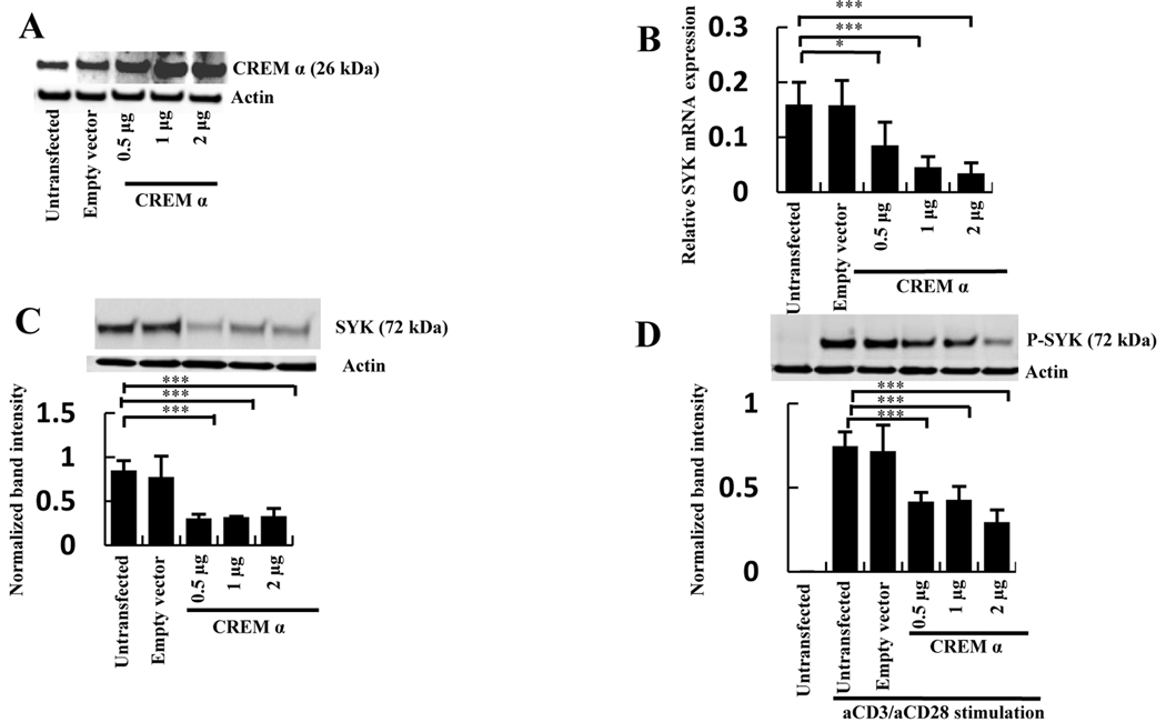
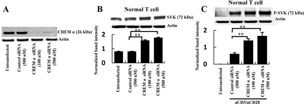
Objective: T cells from patients with systemic lupus erythematosus (SLE) display increased amounts of spleen tyrosine kinase (Syk), which is involved in the aberrant CD3/T cell receptor-mediated signaling process, and increased amounts of CREMα, which suppresses the production of interleukin-2. Syk expression can be suppressed by CREMα. This study was undertaken to investigate why CREMα fails to suppress Syk expression in SLE T cells.

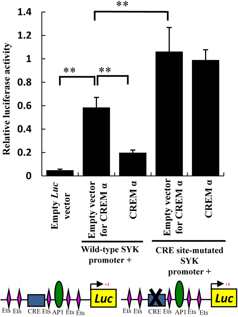
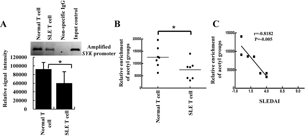
Methods: CREMα was overexpressed in healthy T cells by transfection with CREMα expression vector, and Syk expression and phosphorylation were measured. A newly identified cAMP response element (CRE) site on the SYK promoter was characterized by chromatin immunoprecipitation (ChIP) and electrophoretic mobility shift assay. The CREMα-mediated repression of Syk expression was further evaluated by analyzing SYK promoter activity. T cells from SLE patients and healthy individuals were subjected to ChIP to evaluate CREMα binding and histone H3 acetylation.

Results: Increased CREMα levels suppressed Syk expression by direct binding to a CRE site of the SYK promoter in T cells from healthy individuals but failed to do so in T cells from SLE patients. The failure of CREMα to suppress Syk expression in SLE T cells was due to weaker binding to the CRE site of the SYK promoter compared to healthy T cells because the promoter site is hypoacetylated in SLE T cells and therefore of limited access to transcription factors.

Conclusion: Our findings indicate that epigenetic alteration of the SYK promoter in SLE T cells results in the inability of the transcriptional repressor CREMα to bind and suppress the expression of Syk, resulting in aberrant T cell signaling.


Conflict of interest statement

Conflict statement: None of the authors has an apparent conflict of interest with any company or other entity.

Figures

Figure 1
Figure 1. Overexpression of CREM α downregulates SYK expression
A, T cells were transfected with empty vector or with 0.5, 1 and 2 µg of CREM α expression vector. After 24h post-transfection Western blot was performed with antibodies against CREM α or β-actin. B, T cells were transfected with different concentrations of CREM α expression vector or with an empty vector. SYK mRNA level was detected by quantitative PCR. Relative mRNA expression normalized to 36B4 is shown as mean ± SEM from one of 3 independent experiments. C, T cells were transfected with different concentrations of CREM α expression vector or with an empty vector. After 24h incubation total SYK protein or β-actin were detected by Western blot and represented as mean ± SEM of densitometry scan of 5 different blots. D, T cells were transfected with different concentrations of CREM α expression vector or with an empty vector. The transfected cells were treated with 1 µg anti-CD3/anti-CD28 antibodies and the levels of phosphorylated SYK and β-actin were measured by Western blot. The data presented as mean ± SEM of densitometry scan of 5 independent blots.
Figure 2
Figure 2. Silencing of endogenous CREM α upregulates SYK protein expression and phosphorylation
A, T cells were transfected with 500 nM control siRNA or 100 and 500 nM CREM α-specific siRNA. At 72h post-transfection cells were lysed with RIPA buffer and Western blotted with antibody against CREM α. B, Normal T cells were transfected with 500 nM of control siRNA or 100 and 500 nM CREM α-specific siRNA. Cells were collected after 72h post-transfection in RIPA buffer and Western blotted with antibody against total SYK and β-actin. Data represented as mean ± SEM of densitometry scan of 5 different blots. C, Normal T cells were transfected with 500 nM control siRNA and 100 and 500 nM CREM α-specific siRNA. 1 µg of anti-CD3/anti-CD28 stimulation was given 1h before cell harvesting. Cells were collected after 72h of post-transfection in RIPA buffer and Western blotted with antibody against phosphorylated SYK and β-actin. Data represented as mean ± SEM of densitometry scan of 5 different blots.
Figure 3
Figure 3. CREM α binds on SYK promoter
A, The SYK gene promoter sequence from initiator element to −782. The transcription start site is marked as red; the CRE site on SYK promoter as blue; one AP1 site marked as green; Ets core binding sites as pink. B, Sonicated chromatin from normal T cells was immunoprecipitated by CREM α antibody or a control IgG. Amplified SYK promoter was detected by PCR. C, An oligonucleotude spanning the region of CRE site on SYK promoter was labeled with [γ32P] and incubated with 5 µg T cell nuclear extract. Protein-DNA complexes were separated on a 6% DNA retardation gel and visualized by phosphoimage analyzer. Unlabeled cold competitors (20 to 500 molar excess) were used to displace complexes formed by labeled oligonucleotides. A cold mutated oligonucleotide was used as non specific competitor. One representative experiment out of 3 is shown. D, 2 µg of specific antibody against CREM α or a non-specific IgG were incubated with labeled oligonucleotides and EMSA was performed. E, Mutated CRE site-bearing oligonucleotide was labeled with [γ32P] and EMSA was performed. In a separate reaction a wild-type probe was used. One representative experiment out of 3 is shown.
Figure 4
Figure 4. CREM α overexpression reduces SYK promoter activity
T cells were co-transfected with 2 µg wild-type SYK promoter-luciferase reporter construct, CRE site-mutated SYK promoter-luciferase construct or 500 ng CREM α expression plasmid and 50 ng Renilla plasmid vector as internal control. DNA concentrations were normalized by empty vector. Luciferase readings from cell extract was measured and normalized with Renilla luciferase and the data was plotted as mean ± SEM based on 6 different experiments. Construct cartoon shows upstream cis elements on SYK promoter. Rectangular box indicate CRE site and AP-1 site shown as oval shape. Out of several Ets binding sites on SYK promoter a few are shown as diamond shapes, those are close to CRE and AP-1 sites.
Figure 5
Figure 5. CREM α does not bind to the SYK promoter in SLE T cells
A, Chromatin was prepared from T cells of 5 SLE patients and 5 age- and sex-matched controls and immunoprecipitated with CREM α antibody. One characteristic example of chromatin-immunoprecipitated SYK promoter with CREM α antibody and densitometry scan of 5 separate samples of control and SLE patients are shown as mean ± SEM of relative intensity of immunoprecipitated bands. B, Chromatin was prepared from 7 SLE patients and 7 age- and sex-matched controls and immunoprecipitated with anti-acetyl-histone H3 antibody. Individual immunoprecipitated bands were measured by densitometry and plotted as random scatter. C, Relative enrichment of acetyl group (as depicted on the X axis) was plotted against the respective SLE disease activity index (SLEDAI; Y axis) using Prism graph pad software. Statistical correlation is given as r value.
Figure 6
Figure 6
Model demonstrating CREM α activation by cAMP pathway and subsequent binding on the CRE site of the SYK promoter to result in downregulating SYK expression in normal T cells. In SLE T cells hypoacetylated SYK promoter inhibits CREM α binding on the CRE site and CREM α-mediated suppression of SYK gene expression.

References

    1. Rudd CE. Adaptors and molecular scaffolds in immune cell signaling. Cell. 1999;96(1):5–8. - PubMed
    1. Sada K, Takano T, Yanagi S, Yamamura H. Structure and function of SYK protein-tyrosine kinase. J Biochem. 2001;130(2):177–186. - PubMed
    1. Palacios EH, Weiss A. Function of the Src-family kinases, Lck and Fyn, in T-cell development and activation. Oncogene. 2004;23(48):7990–8000. - PubMed
    1. Chu P, Pardo J, Zhao H, Li CC, Pali E, Shen MM, et al. Systematic identification of regulatory proteins critical for T-cell activation. J Biol. 2003;2(3):21. - PMC - PubMed
    1. Krishnan S, Warke VG, Nambiar MP, Tsokos GC, Farber DL. The FcR gamma subunit and SYK kinase replace the CD3 zeta-chain and ZAP-70 kinase in the TCR signaling complex of human effector CD4 T cells. J Immunol. 2003;170(8):4189–4195. - PubMed

Publication types

MeSH terms