

doi: 10.1002/adma.201102948.
Epub 2011 Sep 26.
Multicore assemblies potentiate magnetic properties of biomagnetic nanoparticles
Affiliations
- PMID: 21953810
- PMCID: PMC3224986
- DOI: 10.1002/adma.201102948
Item in Clipboard
Multicore assemblies potentiate magnetic properties of biomagnetic nanoparticles
Adv Mater.
.
No abstract available
Figures
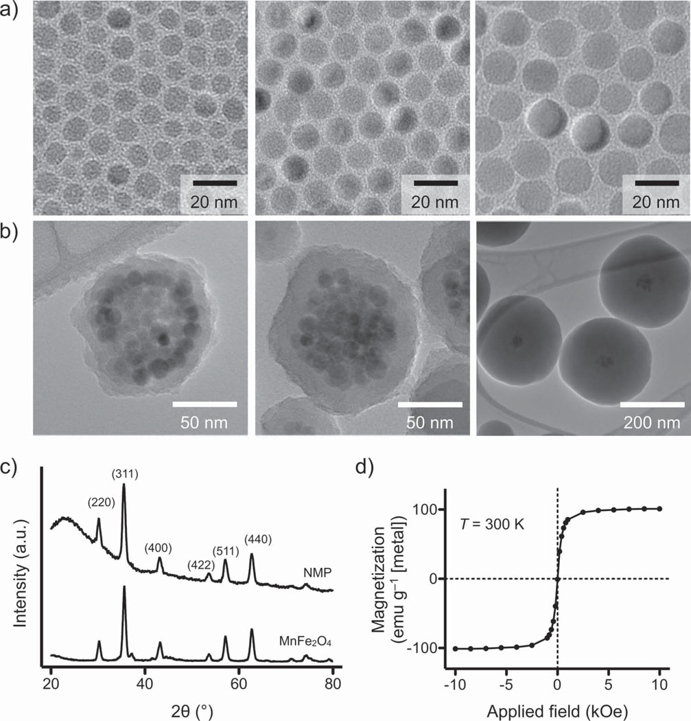
Synthesis and characterization of NMP. a) Mn-doped ferrite crystals were prepared as core MNPs. These core particles were enlarged up to a diameter of 16 nm through seed-mediated growth. All Mn-MNPs had a narrow size distribution and consisted of a single domain. b) NMPs of different sizes were synthesized by encapsulating a cluster of Mn-MNPs (diameter 16 nm) within a silica shell of varying thickness. The estimated number of core Mn-MNPs per NMP was =57. While keeping the same cluster size, the shell thickness was varied from 9 to 140 nm. c) XRD demonstrated that NMPs exhibited identical peak patterns to their unmodified core Mn-MNPs; this confirmed that the cores are preserved during the silica-coating step. The broad background in NMP signal is due to the amorphous nature of the silica shell. d) At T = 300 K, NMPs are superparamagnetic and their saturation magnetization (100 emu g−1 [metal]) is similar to that of 16 nm Mn-MNPs (101 emu g−1 [metal]).
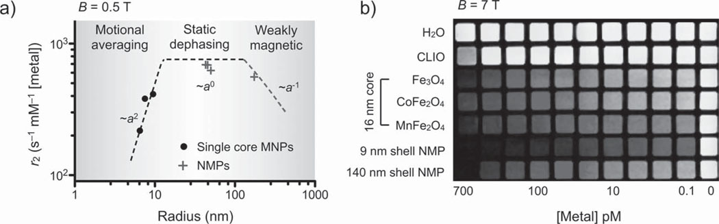
Characterization of r2. a) The r2 values of NMPs and single core Mn-MNPs (at 0.5 T) were compared as a function of particle radius a. While the r2 values of Mn-MNPs increased steeply with increasing particle size (~a2), the r2 values for NMPs initially showed little dependence on particle size (~a0) or showed a gradual decrease at larger sizes (~a−1). Theoretical modeling, based on chemical exchange and static dephasing, was able to accurately describe the observed r2-behavior (dotted lines). Due to their small particle size, Mn-MNPs fell within the motional averaging regime. Most NMPs were in the static dephasing regime with the exception of thick-shelled NMPs, which fell into the weak-magnetic regime. Importantly, the prepared NMPs showed the highest r2 value (695 s−1 mm −1 [metal]), approaching that of the theoretical limit set by the static dephasing model. b) Phantom images (B = 7.0 T) verified the superiority of NMPs as a MR contrasting agent. Note that the Mn-MNPs (used as the NMP cores) were also superior to similarly sized cobalt ferrite (CoFe2 O4) and ferrite (Fe3O4) MNPs, as well as to the conventionally used CLIO (cross-linked iron oxide).
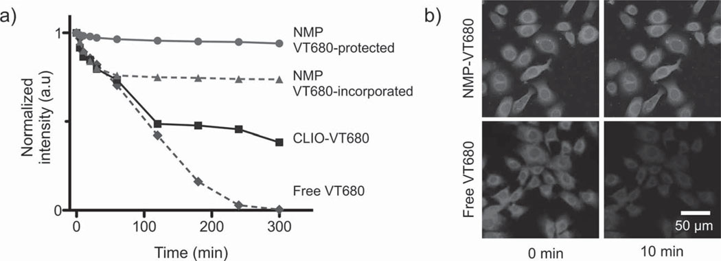
Optical properties of NMPs. a) Photostability of NMPs and other imaging agents were evaluated by monitoring their fluorescence after continuous exposure to ultraviolet light. Signal loss was found to be inversely proportional to the protection level of the dye molecules against the media. NMP-enclosed dye molecules, which had an extra protective silica shell, showed the best stability and least photobleaching (maintaining >97% of their initial intensity). Particles with dyes exposed were less stable, whereas free dyes showed the fastest decline in signal intensity. b) The superb optical stability offered by the NMPs will likely be of enormous benefit to time-lapse high-resolution microscopy, which requires long exposure of samples to an intense light source. Cells targeted with NMP-VT680, for example, showed little change in signal over 1 h under a laser scanning microscope. For cells labeled with free dyes, however, the fluorescence intensity dropped by > 50% in 10 min.
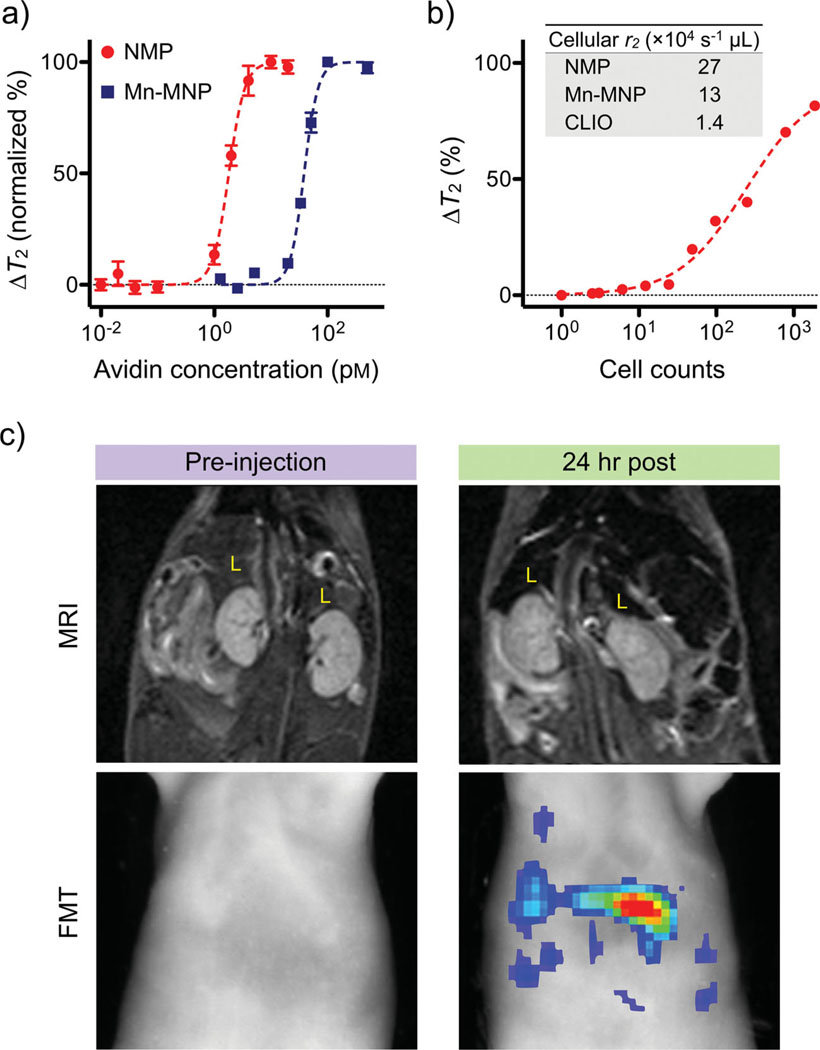
Biological utility of NMPs. a) Small molecule detection was demonstrated using the avidin–biotin system as a model. Biotinylated NMPs were mixed with varying amounts of avidin, which caused aggregation of NMPs and corresponding changes in T2 relaxation times. The high r2 relaxivity of NMPs enabled extremely sensitive detection (≈1 pm of avidin), whereas the detection sensitivity with Mn-MNPs was ≈20-fold lower (20 pm ). b) Cancer cells (SkBr3) targeted with HER2/neu-specific NMPs could be detected in 1 µL sample volumes, and the detection limit was near single-cell level. The inset table compares the cellular relaxivities of different particle preparations. c) NMPs incorporating near-infrared dyes (NMP-VT680) were used as dual in vivo imaging agents. Mice received intravenous injections of NMP-VT680 before undergoing both magnetic resonance imaging (MRI) and fluorescent-mediated tomography FMT). Due to large amounts of phagocytic cells, the liver (L) showed decreased signal intensity with MRI, while under FMT, the liver showed high fluorescent signals.
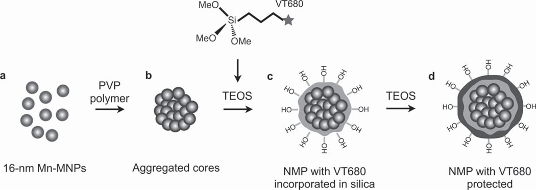
Synthesis route of NMP. a) 16 nm Mn-MNPs were coated with DMSA and dispersed in water. b) DMSA-coated Mn-MNPs were then treated with PVP and induced to form a nanoscale cluster. c) The clusters were encapsulated in silica by polymerizing TEOS via the Stöber process. A near infrared dye (VT680) was co-injected at this point to allow incorporation of the dye into the silica shell. d) The Stöber process was repeated to allow another layer of silica to form, protecting the dye from the external environment.
References
Publication types
MeSH terms
Substances
Grants and funding
LinkOut - more resources
Full Text Sources
Other Literature Sources
