. 2011 Sep;23(9):3482-97.
doi: 10.1105/tpc.111.089581. Epub 2011 Sep 27.

The Arabidopsis Na+/H+ antiporters NHX1 and NHX2 control vacuolar pH and K+ homeostasis to regulate growth, flower development, and reproduction

Affiliations

The Arabidopsis Na+/H+ antiporters NHX1 and NHX2 control vacuolar pH and K+ homeostasis to regulate growth, flower development, and reproduction

Elias Bassil et al. Plant Cell. 2011 Sep.

Erratum in

  • Plant Cell. 2011 Dec;23(12):4526

Abstract

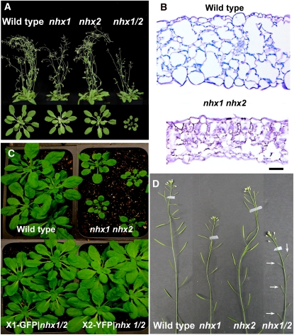
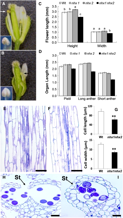
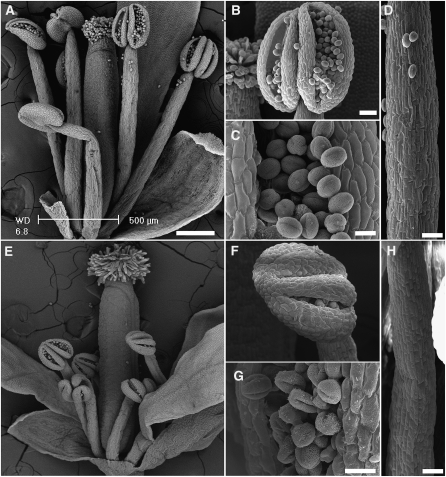
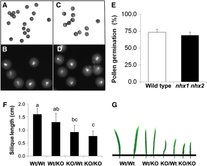
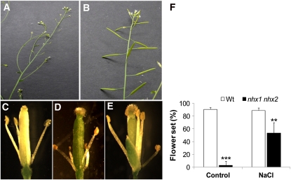
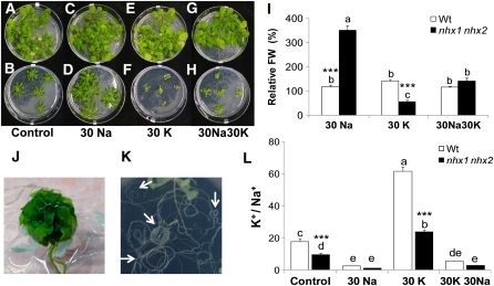
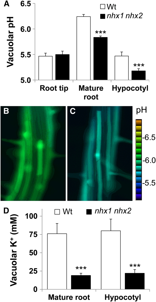
Intracellular Na(+)/H(+) (NHX) antiporters have important roles in cellular pH and Na(+), K(+) homeostasis. The six Arabidopsis thaliana intracellular NHX members are divided into two groups, endosomal (NHX5 and NHX6) and vacuolar (NHX1 to NHX4). Of the vacuolar members, NHX1 has been characterized functionally, but the remaining members have largely unknown roles. Using reverse genetics, we show that, unlike the single knockouts nhx1 or nhx2, the double knockout nhx1 nhx2 had significantly reduced growth, smaller cells, shorter hypocotyls in etiolated seedlings and abnormal stamens in mature flowers. Filaments of nhx1 nhx2 did not elongate and lacked the ability to dehisce and release pollen, resulting in a near lack of silique formation. Pollen viability and germination was not affected. Quantification of vacuolar pH and intravacuolar K(+) concentrations indicated that nhx1 nhx2 vacuoles were more acidic and accumulated only 30% of the wild-type K(+) concentration, highlighting the roles of NHX1 and NHX2 in mediating vacuolar K(+)/H(+) exchange. Growth under added Na(+), but not K(+), partly rescued the flower and growth phenotypes. Our results demonstrate the roles of NHX1 and NHX2 in regulating intravacuolar K(+) and pH, which are essential to cell expansion and flower development.


Figures

Figure 1.
Figure 1.
Subcellular Localization of NHX1 and NHX2. (A) NHX2-associated YFP fluorescence in mature root cells of stably transformed plants expressing 35S-NHX2-YFP. Arrows point to nuclei. (B) Transient expression of Ub10-NHX1-GFP in Arabidopsis cotyledons. (C) to (E) Transient coexpression of Ub10-NHX1-GFP and 35S-NHX2-RFP in Arabidopsis cotyledons. (F) to (H) Transient expression of Ub10-NHX1-GFP in the Ub10-VAMP711-RFP background. (C) and (F) are GFP images; (D) and (G) are RFP images; (E) and (H) are merged images. Arrowheads in (B), (E), and (H) point to the tonoplast and transvacuolar cytoplasmic strands. Bar in (A) = 50 mm; bars in (B) to (H) = 20 mm.
Figure 2.
Figure 2.
Growth and Development of the Single and Double Knockouts of NHX1 and NHX2. (A) Four-week-old plants grown under 16-h days (Top), and 5-week-old plants grown under 8-h days (Bottom). (B) Cross section through the margin of a 4-week-old wild-type leaf (Top) and comparable nhx1 nhx2 knockout leaf (Bottom). Sections were stained with toluidine blue O. (C) The nhx1 nhx2 growth phenotype was rescued by transformation with either 35S-NHX1-GFP or 35S-NHX2-YFP. The wild type and nhx1 nhx2 are included for comparison. Representative images of 4-week-old T1 transformants are shown. (D) Inflorescence stalks of wild-type, nhx1, nhx2, and nhx1 nhx2 plants. Note the lack of siliques in nhx1 nhx2 (arrows). Bar in (B) = 30 μm.
Figure 3.
Figure 3.
Stamens of the Double Knockout nhx1 nhx2 Have Reduced Filament Elongation and Anther Dehiscence. (A) Dissected wild-type flower showing normal filaments and anthers. (B) Dissected nhx1 nhx2 flower showing the short filament (arrows) and nondehiscent anther (inset) phenotypes. (C) Quantification of whole flower size and (D) floral organ size in single and double knockouts of NHX1 and NHX2. Wt, wild type. (E) Longitudinal section of mature wild-type filament. (F) Longitudinal section of mature nhx1 nhx2 filament. (G) Comparison of filament cell length and width. (H) Cross section of a mature wild-type dehiscent anther with a ruptured stomium (St). (I) Cross section of a mature nhx1 nhx2 anther showing the lack of stomium rupture. Different letters in (C) and (D) indicate significant difference using Tukey LSD test (P ≤ 0.05). Values in (G) represent the mean ± sd (n = 10). ** indicate significant difference between genotypes (P ≤ 0.001 and P ≤ 0.01, respectively; Student's t test). Bars in (E), (F), (H), and (I) = 30 μm. [See online article for color version of this figure.]
Figure 4.
Figure 4.
Scanning Electron Micrographs Comparing the Anatomy of the Wild-Type and nhx1 nhx2 Flowers. (A) to (D) Flowers and organs of the wild type. (E) to (F) Flowers and organs of nhx1 nhx2. (A) and (E) Whole flowers; (B) and (F) dehiscent anthers; (C) and (G) pollen grains; (D) and (H) filaments. Bars in (A) and (E) = 200 μm; bars in (B), (F), (D), and (H) = 50 μm; bars in (C) and (G) = 10 μm.
Figure 5.
Figure 5.
Pollen Viability and Germination Is Not Significantly Affected in the nhx1 nhx2 Double Knockout. Wild-type (A) and (B) and nhx1 nhx2 (C) and (D) pollen after 2 h of hydration on gelatin-coated slides. (B) and (D) DAPI staining of hydrated pollen, indicating tricellular pollen. (E) Pollen germination was assessed by scoring germinated pollen grains from an aliquot of pollen incubated in germination media (see Methods) on microscope slides observed within defined areas. A pollen grain was considered germinated when the length of the pollen tube exceeded the diameter of the pollen grain. Approximately 100 pollen grains were counted in each of 25 defined areas. Values are the mean ± sd (n = 25). (F) Quantification of silique length obtained from reciprocal crosses between wild type (wt) and nhx1 nhx2 (KO) (designated as female/male). For each cross, 20 siliques were measured. Values represent the mean ± sd (n = 20). Similar letters indicate no significant difference (P ≤ 0.05) using Tukey LSD test. (G) Picture representation of the data in (F). [See online article for color version of this figure.]
Figure 6.
Figure 6.
The nhx1 nhx2 Flower Phenotype Is Rescued When Plants Are Grown in High Salt. Plants were grown in soil under short days (8 h light) for 4 weeks and then irrigated with 50 mM NaCl followed by 100 mM NaCl 2 d later. An additional 25 mM NaCl was included once a week with irrigation. At 5 weeks, the plants were switched to long days (16 h light) to induce flowering. At 7 weeks, flowers were examined. (A) nhx1 nhx2 inflorescence from plants grown under control conditions showing the characteristic lack of developed siliques. (B) Inflorescence of nhx1 nhx2 when grown in soil supplemented with 100 mM NaCl. (C) Dissected wild-type flower showing fully elongated and dehiscent anthers. (D) Dissected nhx1 nhx2 flower from plants grown without added salt showing short filaments and nondehiscent anthers. (E) Dissected nhx1 nhx2 flower from plants grown under 100 mM NaCl showing fully elongated and dehiscent anthers. (F) Comparison of flower set (%) in wild-type (Wt) and nhx1 nhx2 plants grown in the presence and absence of 100 mM NaCl. The primary inflorescence of each of 10 plants was used for counts. ** and ***, significant difference between genotypes (P ≤ 0.01 and P ≤ 0.001, respectively). Values are the mean ± sd (n = 25 flowers). The experiment was repeated twice. [See online article for color version of this figure.]
Figure 7.
Figure 7.
Growth and Ion Content of the nhx1 nhx2 Double Knockout Depends on K+ and Na+ in Media. Four-week-old wild-type plants (A), (C), (E), and (G) and nhx1 nhx2 (B), (D), (F), and (H) were grown in modified Spalding media containing: (A) and (B) 1 mM K+ and 1 mM Na+ (control), (C) and (D) 1 mM K+ and 30 mM Na+ (30Na), (E) and (F) 30 mM K+ and 1 mM Na+ (30K), and (G) and (H) 30 mM K+ and 30 mM Na+ (30K30Na). (I) Relative shoot fresh weight (FW) (compared with control media of each genotype) of plants shown in (A) to (H). Values are the mean ± sd (n = 25 plants). Similar letters indicate no significant difference (P < 0.05) using Tukey LSD test. ***, significant difference between genotypes (P ≤ 0.01; Student's t test). Wt, wild type. (J) Close-up image of nhx1 nhx2 shoots grown in 30 mM K+. (K) Close-up image of nhx1 nhx2 showing severe root curling in 30 mM K+ but not 30 mM Na+. Arrows point to regions of profuse root curling. (L) Ratio of leaf K+ and Na+ content. Values are the mean ± sd (n = 5). Similar letters indicate no significant difference (P < 0.05) using Tukey LSD test. ***, significant difference between genotypes (P ≤ 0.001, respectively).
Figure 8.
Figure 8.
Vacuoles of nhx1 nhx2 in Root and Hypocotyl Cells Are More Acidic and Contain Less K+ than Comparable Wild-Type Cells. (A) Vacuolar pH in roots and hypocotyl of 4-d etiolated seedlings. Wt, wild type. (B) Ratio images of wild-type or (C) nhx1 nhx2 cells of the mature root zone, indicating lower pH in nhx1 nhx2. The Intensity Modulated Display mode of MetaMorph (Molecular Devices) was used to generate the ratio images and accompanying scale bar. pH was calculated from the fluorescence ratio of confocal images collected in roots and hypocotyls cells of seedlings loaded with the pH-sensitive dye BCECF-AM. After background correction, an integrated pixel intensity value (ImageJ 1.43, National Institutes of Health) was calculated in emission (535 to 550 nm) images after excitation with 488 nm and divided by those acquired when excited by 458 nm to obtain ratio images. Ratio images were used to calculate pH from a calibration curve (see Supplemental Figure 7 online) generated as described in Methods. Error bars are the sd of 35 measurements (i.e., ratio images) representing approximately 15 to 20 cells in at least 10 different seedlings. The experiment was repeated six times. Particular care was taken to use images from similar regions of the root, because a comparison of pH along the root indicated that vacuolar pH is not uniform across all cells along the seedling root (mature root and root tip pH values). (D) The vacuolar K+ concentration is lower in nhx1 nhx2 seedlings than the wild type. Vacuolar K+ was measured as described for BCECF-AM except the dye PBFI-AM was used (see Methods). Values are the mean ± sd (n = 30). ***, significant difference (P ≤ 0.001; Student's t test).

Comment in

References

    1. Aharon G.S., Apse M.P., Duan S., Hua X., Blumwald E. (2003). Characterization of a family of vacuolar Na+/H+ antiporters in Arabidopsis thaliana. Plant Soil 253: 245–256
    1. Alandete-Saez M., Ron M., McCormick S. (2008). GEX3, expressed in the male gametophyte and in the egg cell of Arabidopsis thaliana, is essential for micropylar pollen tube guidance and plays a role during early embryogenesis. Molecular Plant 1: 586–598 - PubMed
    1. Amtmann A., Leigh R. (2010). Ion homeostasis. Abiotic Stress Adaptation in Plants, Pareek A., Sopory S.K., Bohnert H.J., (Dordrecht, The Netherlands: Springer; ), pp. 245–262
    1. Apse M.P., Blumwald E. (2007). Na+ transport in plants. FEBS Lett. 581: 2247–2254 - PubMed
    1. Apse M.P., Sottosanto J.B., Blumwald E. (2003). Vacuolar cation/H+ exchange, ion homeostasis, and leaf development are altered in a T-DNA insertional mutant of AtNHX1, the Arabidopsis vacuolar Na+/H+ antiporter. Plant J. 36: 229–239 - PubMed

Publication types

MeSH terms

LinkOut - more resources