

Conditioned medium from amniotic mesenchymal tissue cells reduces progression of bleomycin-induced lung fibrosis
- PMID: 21954836
- PMCID: PMC3279140
- DOI: 10.3109/14653249.2011.613930
Conditioned medium from amniotic mesenchymal tissue cells reduces progression of bleomycin-induced lung fibrosis
Abstract
Background and aims: We have demonstrated recently that transplantation of placental membrane-derived cells reduces bleomycin-induced lung fibrosis in mice, despite a limited presence of transplanted cells in host lungs. Because placenta-derived cells are known to release factors with potential immunomodulatory and trophic activities, we hypothesized that transplanted cells may promote lung tissue repair via paracrine-acting molecules. To test this hypothesis, we examined whether administration of conditioned medium (CM) generated from human amniotic mesenchymal tissue cells (AMTC) was able to reduce lung fibrosis in this same animal model.
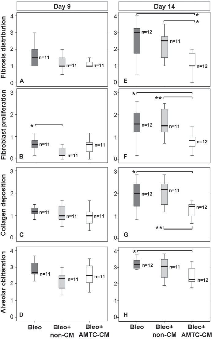
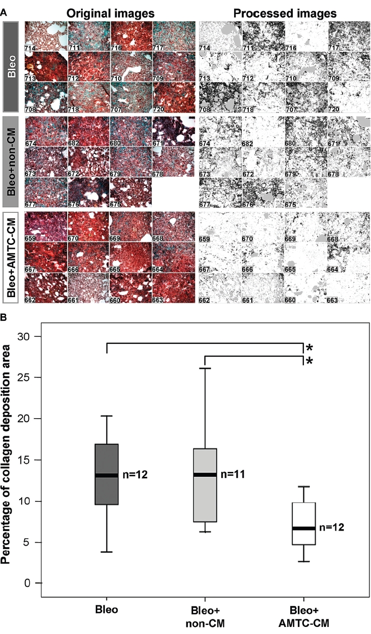
Methods: Bleomycin-challenged mice were either treated with AMTC-CM or control medium, or were left untreated (Bleo group). After 9 and 14 days, the distribution and severity of lung fibrosis were assessed histologically with a scoring system. Collagen deposition was also evaluated by quantitative image analysis.
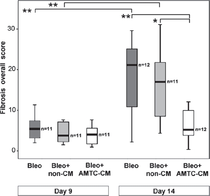
Results: At day 14, lung fibrosis scores in AMTC-CM-treated mice were significantly lower (P < 0.05) compared with mice of the Bleo group, in terms of fibrosis distribution [1.0 (interquartile range, IQR 0.9) versus 3.0 (IQR 1.8)], fibroblast proliferation [0.8 (IQR 0.4) versus 1.6 (IQR 1.0)], collagen deposition [1.4 (IQR 0.5) versus 2.0 (IQR 1.2)] and alveolar obliteration [2.3 (IQR 0.8) versus 3.2 (IQR 0.5)]. No differences were observed between mice of the Bleo group and mice treated with control medium. Quantitative analysis of collagen deposition confirmed these findings. Importantly, AMTC-CM treatment significantly reduced the fibrosis progression between the two observation time-points.
Conclusions: This pilot study supports the notion that AMTC exert anti-fibrotic effects through release of yet unknown soluble factors.
Figures
References
-
- Serrano-Mollar A, Nacher M, Gay-Jordi G, Closa D, Xaubet A, Bulbena O. Intratracheal transplantation of alveolar type II cells reverses bleomycin-induced lung fibrosis. Am J Respir Crit Care Med. 2007;176:1261–8. - PubMed
-
- Germano D, Blyszczuk P, Valaperti A, Kania G, Dirnhofer S, Landmesser U, et al. Prominin-1/CD133+ lung epithelial progenitors protect from bleomycin-induced pulmonary fibrosis. Am J Respir Crit Care Med. 2009;179:939–49. - PubMed
Publication types
MeSH terms
Substances
LinkOut - more resources
Full Text Sources
Other Literature Sources
Medical
