Review
. 2012 Jan 13;25(1):15-34.
doi: 10.1021/tx200339h. Epub 2011 Oct 21.

Biological interactions of graphene-family nanomaterials: an interdisciplinary review

Affiliations
Review

Biological interactions of graphene-family nanomaterials: an interdisciplinary review

Vanesa C Sanchez et al. Chem Res Toxicol. .

Abstract

Graphene is a single-atom thick, two-dimensional sheet of hexagonally arranged carbon atoms isolated from its three-dimensional parent material, graphite. Related materials include few-layer-graphene (FLG), ultrathin graphite, graphene oxide (GO), reduced graphene oxide (rGO), and graphene nanosheets (GNS). This review proposes a systematic nomenclature for this set of Graphene-Family Nanomaterials (GFNs) and discusses specific materials properties relevant for biomolecular and cellular interactions. We discuss several unique modes of interaction between GFNs and nucleic acids, lipid bilayers, and conjugated small molecule drugs and dyes. Some GFNs are produced as dry powders using thermal exfoliation, and in these cases, inhalation is a likely route of human exposure. Some GFNs have aerodynamic sizes that can lead to inhalation and substantial deposition in the human respiratory tract, which may impair lung defense and clearance leading to the formation of granulomas and lung fibrosis. The limited literature on in vitro toxicity suggests that GFNs can be either benign or toxic to cells, and it is hypothesized that the biological response will vary across the material family depending on layer number, lateral size, stiffness, hydrophobicity, surface functionalization, and dose. Generation of reactive oxygen species (ROS) in target cells is a potential mechanism for toxicity, although the extremely high hydrophobic surface area of some GFNs may also lead to significant interactions with membrane lipids leading to direct physical toxicity or adsorption of biological molecules leading to indirect toxicity. Limited in vivo studies demonstrate systemic biodistribution and biopersistence of GFNs following intravenous delivery. Similar to other smooth, continuous, biopersistent implants or foreign bodies, GFNs have the potential to induce foreign body tumors. Long-term adverse health impacts must be considered in the design of GFNs for drug delivery, tissue engineering, and fluorescence-based biomolecular sensing. Future research is needed to explore fundamental biological responses to GFNs including systematic assessment of the physical and chemical material properties related to toxicity. Complete materials characterization and mechanistic toxicity studies are essential for safer design and manufacturing of GFNs in order to optimize biological applications with minimal risks for environmental health and safety.


Figures

Figure 1
Figure 1
Example members of the graphene nanomaterial family and selected properties relevant to colloidal behavior and biological interactions. Graphene oxide sketch adapted with permission from Hamilton .
Figure 2
Figure 2
Surface area stability during aggregation, filtration, or drying for spherical (A) and plate-like (B) particles. Sphere-to-sphere point contact preserves most surface area, but plate alignment destroys surface area if interlayer spaces are inaccessible to adsorbates or surface area measurement probes. This is the case for most GFNs, where interlayer spacing ranges from 0.34 nm – 1 nm, which is too small even for N2 penetration used in BET analysis.
Figure 3
Figure 3
Surface area and bending stiffness for ideal GFNs, calculated from geometry and using 1 TPa for the elastic modulus of a graphene sheet and the area moment of inertia for multilayer materials with interlayer spacing of 0.34 nm.
Figure 4
Figure 4
Adsorption and quenching of dye-labeled DNA on graphene surfaces, and its (1) desorption in presence of c-DNA, (2) exchange with the same DNA in solution, or limited desorption upon increasing temperature up to 95 ° C. Note that the experiments involve GO, but the structure shown is that of graphene, and can be taken to represent the unmodified hydrophobic patches on GO surfaces. Figure adapted from Wu et al. with permission.
Figure 5
Figure 5
DNA cleavage mechanism involving intercalation by GO/Cu2+. Figure from Ren et al. used by permission.
Figure 6
Figure 6
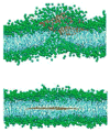
Molecular dynamics simulations showing fusion of lipid-coated monolayer graphene with a lipid bilayer, leading to localization of the sheet in the interleaflet hydrophobic core. Left: intermediate stage of fusion and entry: Right: stable imbedding in hydrophobic core. Figure adapted from Titov et al. and reprinted with permission.
Figure 7
Figure 7
Regional fractional deposition of GFNs in the human respiratory tract. Figures A–C provide particle deposition fractions for particles moving along the polar axis whereas figures D–F provide particle deposition fractions for particles moving perpendicular to the polar axis (A–C) Deposition of 0.5, 5 and 25 μm sized FLGs in the nasopharyngeal, tracheobronchial and alveolar regions respectively; (D–F) Deposition of 0.5, 5 and 25 μm sized FLGs in the nasopharyngeal, tracheobronchial and alveolar regions respectively.
Figure 8
Figure 8
Regional fractional deposition of GFNs in the nasopharyngeal, tracheobronchial and alveolar regions of the human respiratory tract. Lateral dimension ranges from 5 nm to 100 μm. Particles are assumed to be 1 layer thick; layer thickness = 0.34 nm. (A) Particle deposition as the particle moves along the polar axis; and (B) particle deposition as the particle moves perpendicular to the polar axis.
Figure 9
Figure 9
Aerodynamic diameter of various GFNs as a function of layer number. Aerodynamic diameters were determined using two different orientations: (1) particle moves along the polar axis ; and (2) particle moves perpendicular to the polar axis (BLUE).
Figure 10
Figure 10
Size-dependent internalization of FLG by human THP-1 macrophages. Untreated cells (A) or cells exposed to 1μg/ml of carbon black particles (B), multi-walled carbon nanotubes (C), or FLG with increasing lateral size: 550 nm (D), 800 nm (E), 5μm (F) or 25 μm (G). Cells were fixed after 24 hrs of exposure, embedded in plastic, and 0.5 μm sections were stained with toluidine blue for light microscopy. Magnification: 1000X.
Figure 11
Figure 11
Cellular interaction of macrophage with FLG. Following cellular recognition and attachment to FLG with 25 μm of lateral size (A–B), macrophages spread on and wrap the sheets (C–E) without perturbation of their plate-like shape.
Figure 12
Figure 12
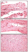
Host response to subcutaneous implants in mice. Subcutaneous implantation of glass/polypropylene transponder identification devices has been shown to induce foreign body sarcomas in heterozygous p53-deficent mice . The initial histopathological reaction (A) to the implant is accumulation of inflammatory cells characterized as macrophages and multinucleated giant cells. The implant becomes surrounded by a dense collagen capsule (B) containing scattered atypical fibroblasts. Atypical cells are large, irregular in shape, and have prominent nuclei. The implant (*) was removed before tissue processing. Foreign body sarcoma (C) composed of densely-packed fibroblast-like cells arranged in whorls. Light microscopy, hematoxylin and eosin stain. Magnification: 200X.

References

    1. Novoselov KS, Geim AK, Morozov SV, Jiang D, Zhang Y, Dubonos SV, Grigorieva IV, Firsov AA. Electric field effect in atomically thin carbon films. Science. 2004;306:666–669. - PubMed
    1. Ruoff R. Graphene: calling all chemists. Nat Nanotechnol. 2008;3:10–11. - PubMed
    1. Stankovich S, Dikin DA, Dommett GH, Kohlhaas KM, Zimney EJ, Stach EA, Piner RD, Nguyen ST, Ruoff RS. Graphene-based composite materials. Nature. 2006;442:282–286. - PubMed
    1. Su FY, You C, He YB, Lv W, Cui W, Jin F, Li B, Yang QH, Kang F. Flexible and planar graphene conductive additives for lithium-ion batteries. J Mater Chem. 2010;20:9644–9650.
    1. Paek SM, Yoo E, Honma I. Enhanced cyclic performance and lithium storage capacity of SnO2/graphene nanoporous electrodes with three-dimensionally delaminated flexible structure. Nano Lett. 2009;9:72–75. - PubMed

Publication types