. 2012 Apr;165(7):2127-39.
doi: 10.1111/j.1476-5381.2011.01702.x.

Thiocolchicoside suppresses osteoclastogenesis induced by RANKL and cancer cells through inhibition of inflammatory pathways: a new use for an old drug

Affiliations

Thiocolchicoside suppresses osteoclastogenesis induced by RANKL and cancer cells through inhibition of inflammatory pathways: a new use for an old drug

Simone Reuter et al. Br J Pharmacol. 2012 Apr.

Abstract

Background and purpose: Most patients with cancer die not because of the tumour in the primary site, but because it has spread to other sites. Common tumours, such as breast, multiple myeloma, and prostate tumours, frequently metastasize to the bone. To search for an inhibitor of cancer-induced bone loss, we investigated the effect of thiocolchicoside, a semi-synthetic colchicoside derived from the plant Gloriosa superba and clinically used as a muscle relaxant, on osteoclastogenesis induced by receptor activator of NF-κB ligand (RANKL) and tumour cells.

Experimental approach: We used RAW 264.7 (murine macrophage) cells, a well-established system for osteoclastogenesis, and evaluated the effect of thiocolchicoside on RANKL-induced NF-κB signalling and osteoclastogenesis as well as on osteoclastogenesis induced by tumour cells.

Key results: Thiocolchicoside suppressed osteoclastogenesis induced by RANKL, and by breast cancer and multiple myeloma cells. Inhibition of the NF-κB pathway was responsible for this effect since the colchicoside inhibited RANKL-induced NF-κB activation, activation of IκB kinase (IKK) and suppressed inhibitor of NF-κBα (IκBα) phosphorylation and degradation, an inhibitor of NF-κB. Furthermore, an inhibitor of the IκBα kinase γ or NF-κB essential modulator, the regulatory component of the IKK complex, demonstrated that the NF-κB signalling pathway is mandatory for osteoclastogenesis induced by RANKL.

Conclusions and implications: Together, these data suggest that thiocolchicoside significantly suppressed osteoclastogenesis induced by RANKL and tumour cells via the NF-κB signalling pathway. Thus, thiocolchicoside, a drug that has been used for almost half a century to treat muscle pain, may also be considered as a new treatment for bone loss.


Figures

Figure 1
Figure 1
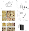
Thiocolchicoside inhibits RANKL-induced osteoclastogenesis. (A) The structure of thiocolchicoside. (B) RAW 264.7 cells (10 × 103 per well) were incubated with 5 nM RANKL, 30 µM thiocolchicoside or both for 3, 4 or 5 days and stained for TRAP expression (red). Magnification, 100× original. (C) RAW 264.7 cells (10 × 103 per well) were incubated with either medium, thiocolchicoside (30 µM) or RANKL (5 nM) alone or with 10, 20 or 30 µM thiocolchicoside plus RANKL for 5 days and then stained for TRAP expression. Magnification, 100× original. (D) Quantification of multinucleated osteoclasts after treatment with medium alone (control; C), with 5 nM RANKL alone or with both RANKL and (10–30 µM) thiocolchicoside for 3, 4 or 5 days. *P < 0.05 and **P < 0.01 indicate level of significance as compared to cells treated with RANKL alone. (E) RAW 264.7 cells (2 × 10³ per 100 µL) were incubated with medium only (Ctrl) or with 10–30 µM thiocolchicoside for 1, 3 or 5 days. Cell proliferation was assessed with the 3-(4,5-dimethylthiazol-2-yl)-2,5-diphenyltetrazolium bromide method. (F) Cells were treated with different concentrations of thiocolchicoside (30–100 µM) and stained with trypan blue. From total number of cells (live + dead), % viable cells were plotted graphically with SD.
Figure 2
Figure 2
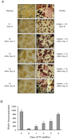
Thiocolchicoside (TC) inhibits RANKL-induced osteoclastogenesis 24 h after stimulation. (A) RAW 264.7 cells (10 × 103 per well) were incubated with RANKL (5 nM) and thiocolchicoside (30 µM) for indicated times and stained for TRAP expression (red). Magnification, 100× original. (B) The number of multinucleated osteoclasts (i.e. those containing three nuclei) was counted. Cells exposed to medium only served as control (C). *P < 0.05 and **P < 0.01 indicate level of significance as compared to cells treated with RANKL alone.
Figure 3
Figure 3
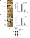
Thiocolchicoside (TC) inhibits osteoclastogenesis induced by cancer cells. (A, left panel) RAW 264.7 cells (10 × 103 per well) were incubated in the presence of MDA-MB-231 cells (1 × 10³ per well), exposed to thiocolchicoside (30 µM) for 5 days, and then stained for TRAP expression. Magnification, 100× original. (A, right panel) RAW 264.7 cells (10 × 103 per well) were incubated either in the presence of MDA-MB-231 cells (1 × 10³ per well) or conditioned medium from MDA-MB-231 cells, exposed to thiocolchicoside (30 µM) for 5 days, and then multinucleated osteoclasts (i.e. those containing three nuclei) were counted. **P < 0.01 indicates level of significance as compared to untreated samples. (B, left panel) RAW 264.7 cells (10 × 103 per well) were incubated in the presence of U266 cells (1 × 10³ per well), exposed to thiocolchicoside (30 µM) for 5 days, and then stained for TRAP expression. Magnification, 100× original. (B, right panel) RAW 264.7 cells (10 × 103 per well) were incubated either in the presence of U266 cells (1 × 10³ per well) or conditioned medium from U266 cells, exposed to thiocolchicoside (30 µM) for 5 days, and then multinucleated osteoclasts (i.e. those containing three nuclei) were counted. **P < 0.01 indicates level of significance as compared to untreated samples. (C) RAW 264.7 cells (2 × 10⁶ per well) were incubated in the presence of conditioned medium from MDA-MB-231 and U266 cells for 24 h and then assessed for NF-κB activity by EMSA.
Figure 4
Figure 4
RANKL induces NF-κB activation and thiocolchicoside inhibits it in a dose- and time-dependent manner. (A) RAW 264.7 cells (1.5 × 10⁶ per well) were incubated with different concentrations of thiocolchicoside for 24 h, treated with 10 nM RANKL for 30 min, and examined for NF-κB activation by EMSA. (B) RAW 264.7 cells (1.5 × 10⁶ per well) were incubated with 50 µM of thiocolchicoside for 24 h and treated with 10 nM RANKL for the indicated times and examined for NF-κB activation by EMSA.
Figure 5
Figure 5
Thiocolchicoside suppresses RANKL-induced IκBα degradation and phosphorylation through inhibition of the IKK activity. (A) RAW 264.7 cells (1.5 × 10⁶ per well) were incubated with 50 µM of thiocolchicoside for 24 h and then treated with 10 nM RANKL for indicated times. Cytoplasmic extracts were examined for IκBα degradation by Western blot using an anti-IκBα antibody. β-Actin was used as a loading control. (B) RAW 264.7 cells (1.5 × 10⁶ per well) were pretreated with thiocolchicoside (50 µM) for 24 h, incubated with ALLN (50 µg·mL−1) for 30 min and treated with RANKL (10 nM) for 15 min. Cytoplasmic extracts were prepared and analysed by Western blot using an anti-phospho-IκBα antibody. β-actin was used as a loading control. (C) RAW 264.7 cells (3 × 10⁶ per well) were pretreated with thiocolchicoside (50 µM) for 24 h and then incubated with RANKL (10 nM) for up to 15 min. Whole-cell extracts were immunoprecipitated using an antibody against IKKα and analysed with an immune complex kinase assay using recombinant GST-IκBα as described in Methods section. To examine the effect of thiocolchicoside on the level of IKK proteins, whole-cell extracts were analysed by Western blot using anti-IKKα and anti-IKKβ antibodies. (D) RAW 264.7 cells (1.5 × 10⁶ per well) were pretreated with thiocolchicoside (50 µM) for 24 h and incubated with RANKL (10 nM) for up to 10 min. Whole-cell extracts were analysed by Western blot using an anti-p-IKKα/β antibody. IKKα was used as a loading control. (E) RAW 264.7 cells (1.5 × 10⁶ per well) were pretreated with thiocolchicoside (50 µM) for 24 h and 48 h and then incubated with RANKL (10 nM) for up to 15 min. Whole-cell extracts were analysed by Western blot with an anti-p-ERK 1/2 antibody. ERK 1/2 was used as a loading control. The numbers below the blot indicate fold activation in comparison to control cells.
Figure 6
Figure 6
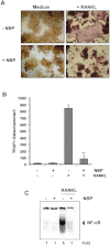
A peptide that targets the NEMO-binding domain inhibits RANKL-induced osteoclastogenesis. (A) RAW 264.7 cells (10 × 103 per well) were pretreated with 100 µM NBP for 2 h, the medium was changed and then RANKL (5 nM) was added for 5 days. Magnification, 100× original. (B) Multinucleated osteoclasts (i.e. those containing three nuclei) were counted. **P < 0.01 indicates level of significance as compared to cells treated with RANKL alone. (C) RAW 264.7 cells (1.5 × 10⁶ per well) were incubated with 100 µM of NBP for 2 h, and then incubated with 10 nM of RANKL for 30 min and examined for NF-κB activation by EMSA.

Comment in

References

    1. Abu-Amer Y, Dowdy SF, Ross FP, Clohisy JC, Teitelbaum SL. TAT fusion proteins containing tyrosine 42-deleted IkappaBalpha arrest osteoclastogenesis. J Biol Chem. 2001;276:30499–30503. - PubMed
    1. Anderson DM, Maraskovsky E, Billingsley WL, Dougall WC, Tometsko ME, Roux ER, et al. A homologue of the TNF receptor and its ligand enhance T-cell growth and dendritic-cell function. Nature. 1997;390:175–179. - PubMed
    1. Bauer DC, Orwoll ES, Fox KM, Vogt TM, Lane NE, Hochberg MC, et al. Aspirin and NSAID use in older women: effect on bone mineral density and fracture risk. Study of Osteoporotic Fractures Research Group. J Bone Miner Res. 1996;11:29–35. - PubMed
    1. Bharti AC, Aggarwal BB. Ranking the role of RANK ligand in apoptosis. Apoptosis. 2004;9:677–690. - PubMed
    1. Bharti AC, Shishodia S, Reuben JM, Weber D, Alexanian R, Raj-Vadhan S, et al. Nuclear factor-kappaB and STAT3 are constitutively active in CD138+ cells derived from multiple myeloma patients, and suppression of these transcription factors leads to apoptosis. Blood. 2004a;103:3175–3184. - PubMed

Publication types

MeSH terms