Hypertrophic lupus erythematosus: the diagnostic utility of CD123 staining
- PMID: 21955314
- PMCID: PMC4103013
- DOI: 10.1111/j.1600-0560.2011.01779.x
Hypertrophic lupus erythematosus: the diagnostic utility of CD123 staining
Abstract
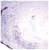
CD123-positive plasmacytoid dendrocytes are prominent in the infiltrate of discoid lupus erythematosus (LE). We hypothesized that these cells would also be present in hypertrophic LE and would aid in the histopathologic distinction from squamous cell carcinoma (SCC) and hypertrophic actinic keratosis (AK). Five cases of hypertrophic LE and 10 cases each of SCC and hypertrophic AK were stained with CD123. A heavy band of CD123-positive cells was present at the epidermal-dermal junction in all cases of hypertrophic LE, and only single or rare scattered clusters of CD123-positive cells were seen in SCC and actinic keratoses. The pattern of CD123 staining can be a useful feature to distinguish hypertrophic LE from SCC and hypertrophic AK.
Copyright © 2011 John Wiley & Sons A/S.
Figures


Similar articles
-
Plasmacytoid dendritic cells in keratoacanthoma and squamous cell carcinoma: A blinded study of CD123 as a diagnostic marker.J Cutan Pathol. 2020 Jan;47(1):17-21. doi: 10.1111/cup.13573. Epub 2019 Sep 1. J Cutan Pathol. 2020. PMID: 31449667
-
Plasmacytoid dendritic cells in hypertrophic discoid lupus erythematosus: an objective evaluation of their diagnostic value.J Cutan Pathol. 2015 Jan;42(1):32-8. doi: 10.1111/cup.12416. Epub 2014 Dec 15. J Cutan Pathol. 2015. PMID: 25367375
-
Differentiating keratoacanthoma from squamous cell carcinoma-In quest of the holy grail.J Cutan Pathol. 2020 Apr;47(4):418-420. doi: 10.1111/cup.13640. Epub 2020 Jan 14. J Cutan Pathol. 2020. PMID: 31893469 Review.
-
Lupus erythematosus mimicking mycosis fungoides: CD123+ plasmacytoid dendritic cells as a useful diagnostic clue.J Cutan Pathol. 2019 Feb;46(2):167-170. doi: 10.1111/cup.13395. Epub 2018 Dec 17. J Cutan Pathol. 2019. PMID: 30430606 No abstract available.
-
[Actinic Keratosis].Laryngorhinootologie. 2015 Jul;94(7):467-79; quiz 480-1. doi: 10.1055/s-0035-1554655. Epub 2015 Jun 30. Laryngorhinootologie. 2015. PMID: 26125293 Review. German.
Cited by
-
Diagnostic utility of plasmacytoid dendritic cells in dermatopathology.Indian J Dermatol Venereol Leprol. 2021 Jan-Feb;87(1):3-13. doi: 10.25259/IJDVL_638_19. Indian J Dermatol Venereol Leprol. 2021. PMID: 33580939 Review.
-
Value of CD123 Immunohistochemistry and Elastic Staining in Differentiating Discoid Lupus Erythematosus from Lichen Planopilaris.Int J Trichology. 2020 Mar-Apr;12(2):62-67. doi: 10.4103/ijt.ijt_32_20. Epub 2020 May 5. Int J Trichology. 2020. PMID: 32684677 Free PMC article.
-
Histiocyte-rich Discoid Lupus Erythematosus: A Peculiar Perifollicular Distribution Histologically Mimicking an Acneiform Disorder.Cureus. 2018 Sep 15;10(9):e3310. doi: 10.7759/cureus.3310. Cureus. 2018. PMID: 32175199 Free PMC article.
-
Solitary verrucous plaque on the chin: hypertrophic discoid lupus erythematosus⋆.An Bras Dermatol. 2025 Mar-Apr;100(2):374-376. doi: 10.1016/j.abd.2024.06.005. Epub 2025 Jan 15. An Bras Dermatol. 2025. PMID: 39818497 Free PMC article. No abstract available.
-
Plasmacytoid Dendritic Cell Marker (CD123) Expression in Scarring and Non-scarring Alopecia.J Cutan Aesthet Surg. 2022 Apr-Jun;15(2):179-182. doi: 10.4103/JCAS.JCAS_126_19. J Cutan Aesthet Surg. 2022. PMID: 35965907 Free PMC article.
References
-
- Daldon PEC, Macedo de Souza E, Cintra ML. Hypertrophic lupus erythematosus: a clinicopathologic study of 14 cases. J Cutan Pathol. 2003;30:443. - PubMed
-
- Jolly HW, Carpenter CL. Multiple keratoacanthomata. Arch Dermatol. 1966;93:348. - PubMed
-
- Fanti PA, Tosti A, Peluso AM, Bonelli U. Multiple keratoacanthoma in discoid lupus erythematosus. J Am Acad Dermatol. 1989;21:809. - PubMed
-
- Uitto J, Santa-Cruz DJ, Eisen AZ, Leone P. Verrucous lesions in patients with discoid lupus erythematosus. Br J Dermatol. 1978;98:507. - PubMed
-
- Perniciaro C, Randle HW, Perry HO. Hypertrophic discoid lupus erythematosus resembling squamous cell carcinoma. Dermatol Surg. 1995;21:255. - PubMed
MeSH terms
Substances
Grants and funding
LinkOut - more resources
Full Text Sources
Other Literature Sources
Medical
Research Materials

