

Hypertrophic lupus erythematosus: the diagnostic utility of CD123 staining
- PMID: 21955314
- PMCID: PMC4103013
- DOI: 10.1111/j.1600-0560.2011.01779.x
Hypertrophic lupus erythematosus: the diagnostic utility of CD123 staining
Abstract
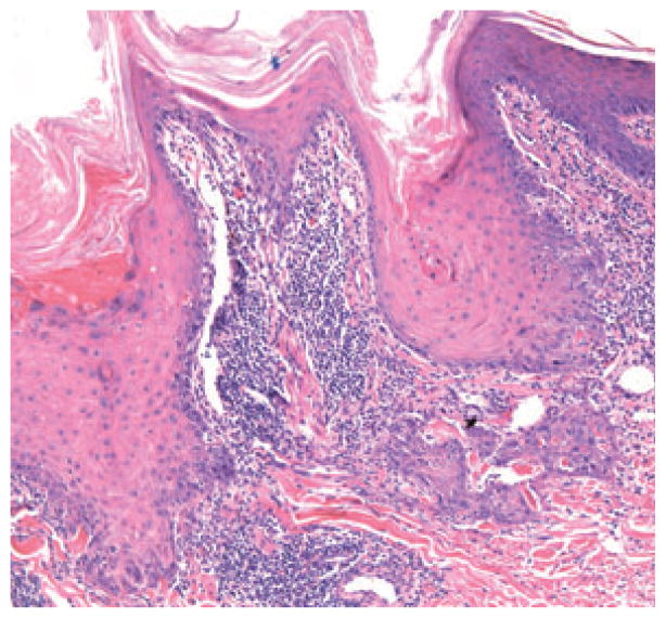
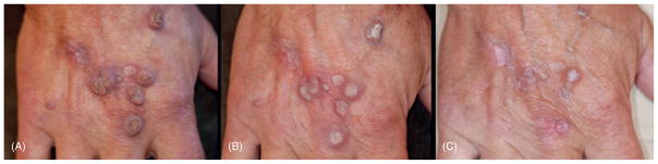
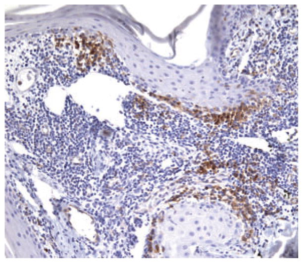
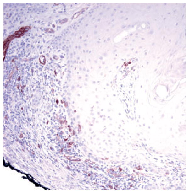
CD123-positive plasmacytoid dendrocytes are prominent in the infiltrate of discoid lupus erythematosus (LE). We hypothesized that these cells would also be present in hypertrophic LE and would aid in the histopathologic distinction from squamous cell carcinoma (SCC) and hypertrophic actinic keratosis (AK). Five cases of hypertrophic LE and 10 cases each of SCC and hypertrophic AK were stained with CD123. A heavy band of CD123-positive cells was present at the epidermal-dermal junction in all cases of hypertrophic LE, and only single or rare scattered clusters of CD123-positive cells were seen in SCC and actinic keratoses. The pattern of CD123 staining can be a useful feature to distinguish hypertrophic LE from SCC and hypertrophic AK.
Copyright © 2011 John Wiley & Sons A/S.
Figures
References
-
- Daldon PEC, Macedo de Souza E, Cintra ML. Hypertrophic lupus erythematosus: a clinicopathologic study of 14 cases. J Cutan Pathol. 2003;30:443. - PubMed
-
- Jolly HW, Carpenter CL. Multiple keratoacanthomata. Arch Dermatol. 1966;93:348. - PubMed
-
- Fanti PA, Tosti A, Peluso AM, Bonelli U. Multiple keratoacanthoma in discoid lupus erythematosus. J Am Acad Dermatol. 1989;21:809. - PubMed
-
- Uitto J, Santa-Cruz DJ, Eisen AZ, Leone P. Verrucous lesions in patients with discoid lupus erythematosus. Br J Dermatol. 1978;98:507. - PubMed
-
- Perniciaro C, Randle HW, Perry HO. Hypertrophic discoid lupus erythematosus resembling squamous cell carcinoma. Dermatol Surg. 1995;21:255. - PubMed
MeSH terms
Substances
Grants and funding
LinkOut - more resources
Full Text Sources
Other Literature Sources
Medical
Research Materials
