. 2011 Sep 28:8:78.
doi: 10.1186/1742-4690-8-78.

Lack of adaptation to human tetherin in HIV-1 group O and P

Affiliations

Lack of adaptation to human tetherin in HIV-1 group O and P

Su Jung Yang et al. Retrovirology. .

Abstract

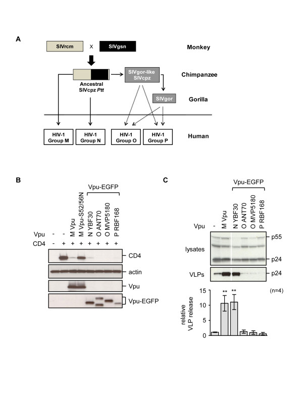
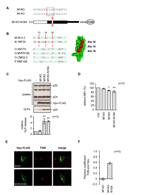
Background: HIV-1 viruses are categorized into four distinct groups: M, N, O and P. Despite the same genomic organization, only the group M viruses are responsible for the world-wide pandemic of AIDS, suggesting better adaptation to human hosts. Previously, it has been reported that the group M Vpu protein is capable of both down-modulating CD4 and counteracting BST-2/tetherin restriction, while the group O Vpu cannot antagonize tetherin. This led us to investigate if group O, and the related group P viruses, possess functional anti-tetherin activities in Vpu or another viral protein, and to further map the residues required for group M Vpu to counteract human tetherin.

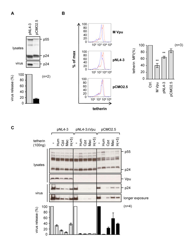
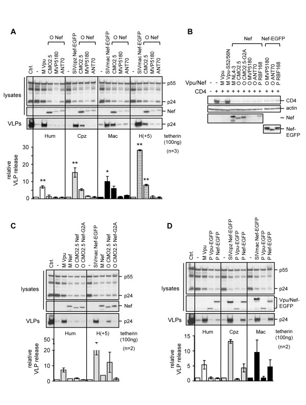
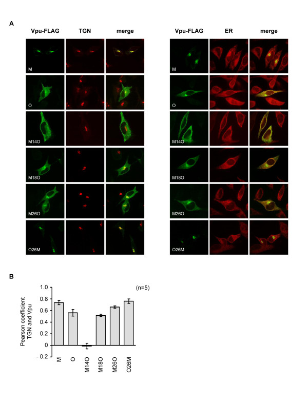
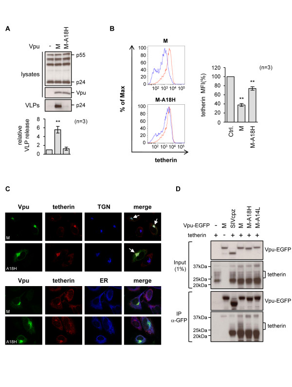
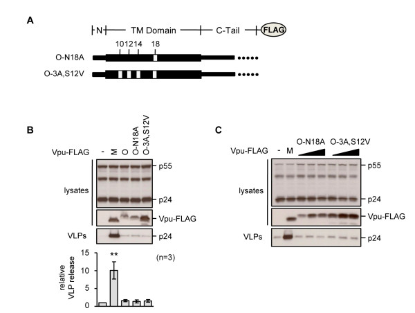
Results: We found a lack of activity against human tetherin for both the Vpu and Nef proteins from group O and P viruses. Furthermore, we found no evidence of anti-human tetherin activity in a fully infectious group O proviral clone, ruling out the possibility of an alternative anti-tetherin factor in this virus. Interestingly, an activity against primate tetherins was retained in the Nef proteins from both a group O and a group P virus. By making chimeras between a functional group M and non-functional group O Vpu protein, we were able to map the first 18 amino acids of group M Vpu as playing an essential role in the ability of the protein to antagonize human tetherin. We further demonstrated the importance of residue alanine-18 for the group M Vpu activity. This residue lies on a diagonal face of conserved alanines in the TM domain of the protein, and is necessary for specific Vpu-tetherin interactions.

Conclusions: The absence of human specific anti-tetherin activities in HIV-1 group O and P suggests a failure of these viruses to adapt to human hosts, which may have limited their spread.


Figures

Figure 1
Figure 1
Anti-tetherin activities of Vpu proteins from major HIV-1 groups. (A) Origins of the four major groups of HIV-1. Solid arrows represent established transmissions, while broken arrows are more speculative events. (B) Ability of Vpu or Vpu-EGFP constructs to degrade CD4, examined by co-transfection of HeLa cells with a CD4 expression plasmid and the indicated Vpu plasmid. Western blots of cell lysates probed with the indicated antibodies are shown. The Vpu- constructs are described by both the HIV-1 group letter and the virus strain. As controls we included a group M Vpu from isolate NL4-3, and its S52/56N mutant that is unable to degrade CD4 [65]. (C) HIV-1 VLP release from tetherin-positive HeLa cells was measured by co-transfection of pHIV-1-pack (expresses HIV-1 Gag-Pol, Rev) in the absence (-) or presence of the indicated Vpu plasmids. VLP release was measured as the ratio of p24-reacting bands in the supernatants versus cell lysates following Western blot analysis, and made relative to the baseline level in the absence of Vpu, for n = 4 independent experiments. Statistical significance is indicated as p < 0.01 (**).
Figure 2
Figure 2
Anti-tetherin activity in group O proviral clone pCMO2.5. (A) Five μg of group M (pNL4-3) or group O (pCMO2.5) proviral clones were transfected into HeLa cells, and cell lysates and supernatants harvested and analyzed by Western blotting with an anti-p24 antibody. The percent virus release was calculated as the ratio of p24-reacting bands in the supernatants relative to the cell lysates, and normalized to 100% for the virus release from pNL4-3, for n = 2 independent experiments. (B) HeLa cells were transfected with 500 ng of a GFP expression plasmid alone (red), or together with 2 μg of either an expression plasmid for group M Vpu, or 5 μg of the proviral clones pNL4-3 or pCMO2.5 (blue). Cells were stained with an anti-tetherin antibody and analyzed for cell surface tetherin expression by FACS. The histograms show relative cell numbers (% of maximum) vs. tetherin expression (fluorescence intensity of APC) in cells gated for GFP expression; graph shows mean MFI in GFP-positive populations for n = 3 independent experiments, p < 0.01 (**). (C) Human (Hum), chimpanzee (Cpz), macaque (Mac), or a chimeric human tetherin, H(+5), containing an insert from Cpz-tetherin in the cytoplasmic tail, were transiently expressed in 293A cells, together with proviral clones pNL4-3, pNL4-3ΔVpu or pCMO2.5. The percent virus release was calculated as described above and made relative to the no tetherin control for each virus, for n = 4 independent experiments. The Vpu antisera used does not cross-react with the group O protein.
Figure 3
Figure 3
Anti-tetherin activities in group O and P Nef proteins. (A) Anti-tetherin activity of group O Nef proteins against the indicated tetherins was examined in 293A cells. Graph shows VLP release in the presence of indicated tetherins and Vpu or Nef proteins relative to the baseline levels of release from the tetherin alone controls (-), for n = 3 independent experiments. Group M Vpu, SIVcpz Nef-EGFP and SIVmac239 Nef-EGFP proteins were included as positive controls. Nef proteins were detected using antiserum raised against group M Nef protein that cross-reacts with group O proteins but not SIVmac Nef. Statistical significance is indicated as p < 0.05 (*) or p < 0.01 (**). (B) Human CD4 expression plasmid (1 μg) was transfected into 293A cells, together with 1 μg of the indicated Vpu or Nef plasmids. Group M Vpu and the defective Vpu-S52/56N mutant were included as positive and negative controls for CD4 degradation, respectively. Untagged Nef proteins were probed using anti-group M Nef antiserum and GFP-tagged Nef proteins were detected using anti-GFP antibody. (C) Activity of CMO2.5 Nef and a myristoylation site mutant (CMO2.5 Nef-G2A) against human and H(+5) tetherin, in 293A cells. Group M Vpu and SIVmac Nef were included as positive controls and group M Nef was included as a negative control. (D) Effect of group P Vpu or Nef proteins on HIV-1 VLP release in the presence of different tetherins, measured in 293A cells, as previously described, for n = 2 independent experiments. Vpu and Nef expression was detected using anti-GFP antibody.
Figure 4
Figure 4
Characterization of chimeric M-O Vpu proteins. (A) Schematic (not to scale) of major domains in FLAG-tagged chimeric Vpu proteins formed between the functional group M (NL4-3, grey) and non-functional group O (ANT70, black) proteins. Numbers in name indicate junction site and refer to the group M residues. (B) Activity of M-O chimeric Vpu-FLAG proteins against human tetherin in HeLa cells. Relative VLP release was calculated as described previously and is shown for n = 3 independent experiments, p < 0.01 (**). Expression of Vpu-FLAG proteins was confirmed using an anti-FLAG antibody. (C) Vpu-FLAG proteins O and O26M are expressed at lower levels than other Vpu constructs, so increasing amounts of the plasmids were transfected into HeLa cells (range 2 to 6 μg), to confirm that their lack of anti-tetherin activity was not simply due to lower levels of expression. As a control, 2 μg of group M Vpu-FLAG was transfected. (D) Ability of chimeric M-O Vpu-FLAG proteins to remove tetherin from the surface of HeLa cells. Cells were co-transfected with 2 μg of indicated Vpu plasmid and 500 ng of GFP expression plasmid and MFI calculated in the GFP-positive population. Graph shows mean MFI for n = 3 independent experiments, p < 0.01 (**). (E) 293T cells were transfected with HA-tagged tetherin alone (500 ng) or together with the indicated Vpu-FLAG expression plasmids (1 μg), except O and O26M (2 μg). Immunoprecipitation (IP) was performed using anti-HA MicroBeads, followed by Western blot analysis of both input lysates (1%) and immunoprecipitates, using anti-FLAG and anti-tetherin antibodies.
Figure 5
Figure 5
Subcellular localization of chimeric M-O Vpu proteins. (A) Subcellular localization of Vpu chimeras in HeLa cells, transfected with the indicated Vpu-FLAG chimeras and stained with antiserum against FLAG (green), and TGN (left) or ER (right) markers (red). (B) The degree of co-localization of Vpu proteins with the TGN marker was calculated using the Pearson coefficient.
Figure 6
Figure 6
Role of Alanine-18 in tetherin antagonism by M-O chimeric Vpu proteins. (A) Schematic of TM domains from M14O and M18O, highlighting location of alanine-18, and configuration of M14O-N18A. (B) Sequence alignment of Vpu TM domains from indicated viruses, with numbering based on group M protein. Alanine residues that are conserved in the functional group M and N Vpu proteins, but are absent in the non-functional group O and P proteins, are labeled in red; non-aromatic hydrophobic residues are labeled in green. Also shown is the 3-D structure of the group M Vpu TM domain (residues 7 to 25 from isolate BH10) [57], created using PyMOL software (Schrödinger LLC), with the conserved alanine residues highlighted in red. (C) Effects of indicated Vpu proteins on HIV-1 VLP release from HeLa cells, measured as previously described, p < 0.01 (**). (D) Effects of indicated Vpu proteins on cell surface tetherin in HeLa cell, measured as previously described, p < 0.05 (*) or p < 0.01 (**). (E) Subcellular localization of M14O and M14O-N18A proteins in HeLa cells, detected by confocal microscopy. Vpu proteins were visualized using anti-FLAG antibody (green), and the TGN (red) was detected with specific antisera. (F) The degree of co-localization of Vpu proteins with the TGN marker was calculated using the Pearson coefficient.
Figure 7
Figure 7
Alanine-18 is important for group M Vpu anti-tetherin activity. (A) Effect of group M Vpu and the A18H mutant on VLP release from HeLa cells, measured as previously described, p < 0.01 (**). (B) Effect of Vpu on tetherin expression on cell surface of HeLa cells, in the absence (red) or presence (blue) of Vpu, examined as previously described. p < 0.01 (**) (C) Subcellular localization of indicated Vpu constructs in HeLa cells and their effects on tetherin distribution. TGN (top) and ER (bottom) markers are included. Arrows indicate cells expressing Vpu that redistributed tetherin to the TGN. (D) 293T cells were transfected with tetherin alone (500 ng) or together with the indicated Vpu-EGFP expression plasmids (5 μg), where M is the Vpu protein from group M strain HXB2. SIVcpz Vpu-EGFP, which is not active against human tetherin, and a Vpu mutant, A14L, were included as negative controls. Immunoprecipitation (IP) was performed using anti-GFP MicroBeads, followed by Western blot analysis of both input lysates (1%) and immunoprecipitates, using either anti-GFP or anti-tetherin antibodies. Mature glycosylated forms of tetherin run between 25 and 37 kDa (bracketed) and interact specifically with wild-type group M Vpu, while an immature faster running species that is also present in the transfected cells is non-specifically pulled down by all Vpu proteins.
Figure 8
Figure 8
Residues from group M Vpu are not sufficient to confer anti-tetherin activity to group O Vpu. (A) Schematic of Vpu-FLAG proteins based on group O Vpu, with group M substitutions at positions 10, 12, 14 and 18. (B) Ability of indicated Vpu proteins to increase HIV-1 VLP release from HeLa cells, measured as previously described, p < 0.01 (**). (C) Effect of transfecting increasing amounts of plasmids O-N18A and O-3A,S12V (range 2 to 6 μg) on VLP release from HeLa cells.

Similar articles

Cited by

References

    1. Neil SJ, Zang T, Bieniasz PD. Tetherin inhibits retrovirus release and is antagonized by HIV-1 Vpu. Nature. 2008;451:425–430. doi: 10.1038/nature06553. - DOI - PubMed
    1. Van Damme N, Goff D, Katsura C, Jorgenson RL, Mitchell R, Johnson MC, Stephens EB, Guatelli JC. The interferon-induced protein BST-2 restricts HIV-1 release and is downregulated from the cell surface by the viral Vpu protein. Cell Host Microbe. 2008;3:245–252. doi: 10.1016/j.chom.2008.03.001. - DOI - PMC - PubMed
    1. Sauter D, Schindler M, Specht A, Landford WN, Münch J, Kim KA, Votteler J, Schubert U, Bibollet-Ruche F, Keele BF, Takehisa J, Ogando Y, Ochsenbauer C, Kappes JC, Ayouba A, Peeters M, Learn GH, Shaw G, Sharp PM, Bieniasz PD, Hahn BH, Hatziioannou T, Kirchhoff F. Tetherin-driven adaptation of Vpu and Nef function and the evolution of pandemic and nonpandemic HIV-1 strains. Cell Host Microbe. 2009;6:409–421. doi: 10.1016/j.chom.2009.10.004. - DOI - PMC - PubMed
    1. Jia B, Serra-Moreno R, Neidermyer W, Rahmberg A, Mackey J, Fofana IB, Johnson WE, Westmoreland S, Evans DT. Species-specific activity of SIV Nef and HIV-1 Vpu in overcoming restriction by tetherin/BST2. PLoS Pathog. 2009;5:e1000429. doi: 10.1371/journal.ppat.1000429. - DOI - PMC - PubMed
    1. Zhang F, Wilson SJ, Landford WC, Virgen B, Gregory D, Johnson MC, Munch J, Kirchhoff F, Bieniasz PD, Hatziioannou T. Nef proteins from simian immunodeficiency viruses are tetherin antagonists. Cell Host Microbe. 2009;6:54–67. doi: 10.1016/j.chom.2009.05.008. - DOI - PMC - PubMed

Publication types

MeSH terms

Substances