

Ribonucleotide reductase inhibition by metal complexes of Triapine (3-aminopyridine-2-carboxaldehyde thiosemicarbazone): a combined experimental and theoretical study
- PMID: 21955844
- PMCID: PMC3374975
- DOI: 10.1016/j.jinorgbio.2011.07.003
Ribonucleotide reductase inhibition by metal complexes of Triapine (3-aminopyridine-2-carboxaldehyde thiosemicarbazone): a combined experimental and theoretical study
Abstract
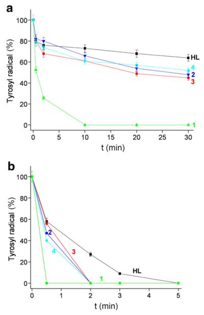
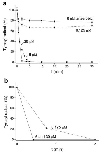
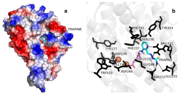
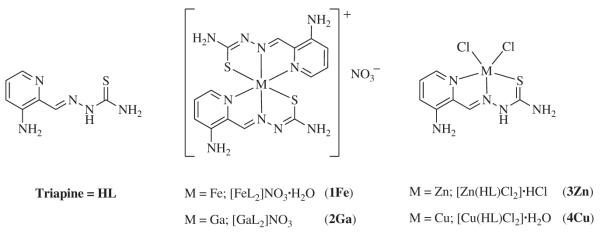
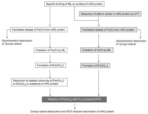
Triapine (3-aminopyridine-2-carboxaldehyde thiosemicarbazone, 3-AP) is currently the most promising chemotherapeutic compound among the class of α-N-heterocyclic thiosemicarbazones. Here we report further insights into the mechanism(s) of anticancer drug activity and inhibition of mouse ribonucleotide reductase (RNR) by Triapine. In addition to the metal-free ligand, its iron(III), gallium(III), zinc(II) and copper(II) complexes were studied, aiming to correlate their cytotoxic activities with their effects on the diferric/tyrosyl radical center of the RNR enzyme in vitro. In this study we propose for the first time a potential specific binding pocket for Triapine on the surface of the mouse R2 RNR protein. In our mechanistic model, interaction with Triapine results in the labilization of the diferric center in the R2 protein. Subsequently the Triapine molecules act as iron chelators. In the absence of external reductants, and in presence of the mouse R2 RNR protein, catalytic amounts of the iron(III)-Triapine are reduced to the iron(II)-Triapine complex. In the presence of an external reductant (dithiothreitol), stoichiometric amounts of the potently reactive iron(II)-Triapine complex are formed. Formation of the iron(II)-Triapine complex, as the essential part of the reaction outcome, promotes further reactions with molecular oxygen, which give rise to reactive oxygen species (ROS) and thereby damage the RNR enzyme. Triapine affects the diferric center of the mouse R2 protein and, unlike hydroxyurea, is not a potent reductant, not likely to act directly on the tyrosyl radical.
Copyright © 2011. Published by Elsevier Inc.
Figures
Similar articles
-
Activity and electrochemical properties: iron complexes of the anticancer drug triapine and its analogs.J Biol Inorg Chem. 2019 Aug;24(5):621-632. doi: 10.1007/s00775-019-01675-0. Epub 2019 Jun 27. J Biol Inorg Chem. 2019. PMID: 31250199 Free PMC article.
-
Impact of metal coordination on cytotoxicity of 3-aminopyridine-2-carboxaldehyde thiosemicarbazone (triapine) and novel insights into terminal dimethylation.J Med Chem. 2009 Aug 27;52(16):5032-43. doi: 10.1021/jm900528d. J Med Chem. 2009. PMID: 19637923
-
Triapine Derivatives Act as Copper Delivery Vehicles to Induce Deadly Metal Overload in Cancer Cells.Biomolecules. 2020 Sep 19;10(9):1336. doi: 10.3390/biom10091336. Biomolecules. 2020. PMID: 32961653 Free PMC article.
-
Iron-targeting antitumor activity of gallium compounds and novel insights into triapine(®)-metal complexes.Antioxid Redox Signal. 2013 Mar 10;18(8):956-72. doi: 10.1089/ars.2012.4880. Epub 2012 Oct 3. Antioxid Redox Signal. 2013. PMID: 22900955 Free PMC article. Review.
-
Syntheses and antitumor activities of potent inhibitors of ribonucleotide reductase: 3-amino-4-methylpyridine-2-carboxaldehyde-thiosemicarba-zone (3-AMP), 3-amino-pyridine-2-carboxaldehyde-thiosemicarbazone (3-AP) and its water-soluble prodrugs.Curr Med Chem. 2001 Feb;8(2):121-33. doi: 10.2174/0929867013373741. Curr Med Chem. 2001. PMID: 11172670 Review.
Cited by
-
Mechanistic insights on the mode of action of an antiproliferative thiosemicarbazone-nickel complex revealed by an integrated chemogenomic profiling study.Sci Rep. 2020 Jun 29;10(1):10524. doi: 10.1038/s41598-020-67439-y. Sci Rep. 2020. PMID: 32601343 Free PMC article.
-
Therapeutic Mechanisms of Treatment in Cervical and Vaginal Cancer.Oncol Hematol Rev. 2012 Spring;8(1):55-60. Oncol Hematol Rev. 2012. PMID: 22943045 Free PMC article.
-
A small-molecule blocking ribonucleotide reductase holoenzyme formation inhibits cancer cell growth and overcomes drug resistance.Cancer Res. 2013 Nov 1;73(21):6484-93. doi: 10.1158/0008-5472.CAN-13-1094. Epub 2013 Sep 26. Cancer Res. 2013. PMID: 24072748 Free PMC article.
-
Copper(II) Thiosemicarbazone Complexes and Their Proligands upon UVA Irradiation: An EPR and Spectrophotometric Steady-State Study.Molecules. 2018 Mar 21;23(4):721. doi: 10.3390/molecules23040721. Molecules. 2018. PMID: 29561827 Free PMC article.
-
Intracellular reduction/activation of a disulfide switch in thiosemicarbazone iron chelators.Metallomics. 2014 Oct;6(10):1905-12. doi: 10.1039/c4mt00153b. Epub 2014 Aug 7. Metallomics. 2014. PMID: 25100578 Free PMC article.
References
-
- Thelander L, Reichard P. Ann. Rev. Biochem. 1979;48:133–158. - PubMed
-
- Thelander L, Gräslund A. J. Biol. Chem. 1983;258:4063–4066. - PubMed
-
- Moore EC, Sartorelli AC. In: Inhibitors of Ribonucleoside Diphosphate Reductase Activity. Cory GJ, Cory AH, editors. Pergamon Press; Oxford: 1989. pp. 203–215.
-
- Chaston TB, Lovejoy DB, Watts RN, Richardson DR. Clin. Cancer Res. 2003;9:402–414. - PubMed
-
- Yu Y, Kalinowski DS, Kovacevic Z, Siafakas AR, Jansson PJ, Stefani C, Lovejoy DB, Sharpe PC, Bernhardt PV, Richardson DR. J. Med. Chem. 2009;52:5271–5294. - PubMed
Publication types
MeSH terms
Substances
Grants and funding
LinkOut - more resources
Full Text Sources
Other Literature Sources
