. 2011 Sep 28;12(9):R96.
doi: 10.1186/gb-2011-12-9-r96.

First somatic mutation of E2F1 in a critical DNA binding residue discovered in well-differentiated papillary mesothelioma of the peritoneum

Affiliations

First somatic mutation of E2F1 in a critical DNA binding residue discovered in well-differentiated papillary mesothelioma of the peritoneum

Willie Yu et al. Genome Biol. .

Abstract

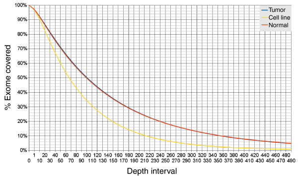
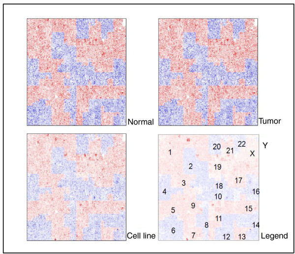
Background: Well differentiated papillary mesothelioma of the peritoneum (WDPMP) is a rare variant of epithelial mesothelioma of low malignancy potential, usually found in women with no history of asbestos exposure. In this study, we perform the first exome sequencing of WDPMP.

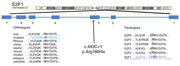
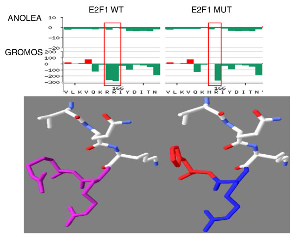
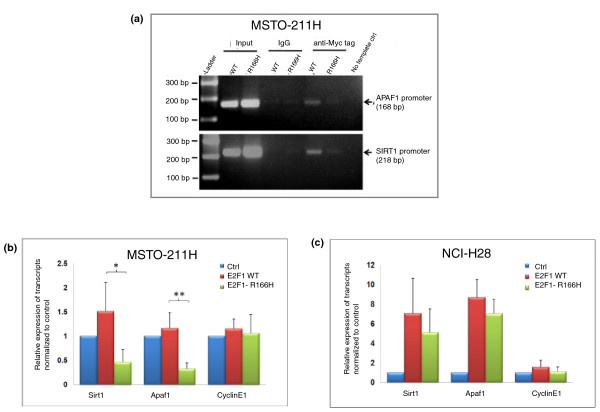
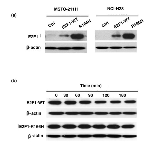
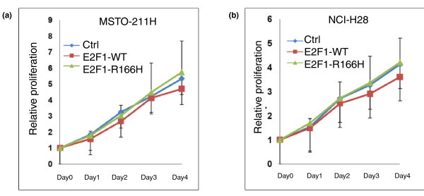
Results: WDPMP exome sequencing reveals the first somatic mutation of E2F1, R166H, to be identified in human cancer. The location is in the evolutionarily conserved DNA binding domain and computationally predicted to be mutated in the critical contact point between E2F1 and its DNA target. We show that the R166H mutation abrogates E2F1's DNA binding ability and is associated with reduced activation of E2F1 downstream target genes. Mutant E2F1 proteins are also observed in higher quantities when compared with wild-type E2F1 protein levels and the mutant protein's resistance to degradation was found to be the cause of its accumulation within mutant over-expressing cells. Cells over-expressing wild-type E2F1 show decreased proliferation compared to mutant over-expressing cells, but cell proliferation rates of mutant over-expressing cells were comparable to cells over-expressing the empty vector.

Conclusions: The R166H mutation in E2F1 is shown to have a deleterious effect on its DNA binding ability as well as increasing its stability and subsequent accumulation in R166H mutant cells. Based on the results, two compatible theories can be formed: R166H mutation appears to allow for protein over-expression while minimizing the apoptotic consequence and the R166H mutation may behave similarly to SV40 large T antigen, inhibiting tumor suppressive functions of retinoblastoma protein 1.


Figures

Figure 1
Figure 1
Cumulative WDPMP exome coverage for the tumor, normal sample and tumor derived cell line. Cumulative exome coverage curve for the tumor (blue), normal sample (orange) and cell line (yellow) is generated by plotting the percentage of the exome represented by different read depths where read depth is defined as the number of individual 75-bp sequenced reads mapped to a particular exome position. The 'fat tail' of the graph indicates a bias in the capturing technology as small sections of the exome are over-represented.
Figure 2
Figure 2
Compact representation of the WDPMP exome using Hilbert plots. Instead of linearly plotting the sequencing depth versus the exome DNA string, HilbertVis [11] computationally wraps the DNA string in a fractal manner onto a two-dimensional grid of pre-determined size and represents the coverage depth via a heat map similar to gene expression data. Red and blue color heat mapping is used to demarcate the borders of each chromosome.
Figure 3
Figure 3
Location and conservation analysis of E2F1 R166H. E2F1 genomic location, exon location of c.493 c > Y mutation and results of E2F1 mutation validation and conservation analysis. Top: the chromosomal location of E2F1 and the location of its exons. The exon numbering indicates E2F1 is located on the reverse strand and the c.493C > Y mutation is location on exon 3, which translates to a p.Arg166His residue mutation. E2F1 orthologue conservation analysis was performed using the SNP Analysis function of SNPs3D [15] with the E2F1 mutated protein sequence shown in light blue (bottom left). The arginine-arginine conservation across diverse species is shown with the histidine mutation highlighted in red and its arginine partner highlighted in blue. E2F1 paralogue conservation analysis was performed using CLUSTALW [14] at default settings (bottom right). The E2F1 mutated sequence is shown in light blue and underlined with the histidine mutation shown in red and its partner arginine shown in blue. Again the arginine-arginine conservation across the E2F family is clearly shown.
Figure 4
Figure 4
Visualization of the p.Arg166His mutation in E2F1. Top: the E2F4 crystal structure [PDB:1CF7] showing the location of the p.Arg166His mutation. The brown double helix is the DNA binding motif with green colored guanine nucleotides representing binding targets of Arg182 and Arg183 of the DP2 protein and yellow colored guanine nucleotides representing binding targets of Arg166 and Arg165 of the E2F protein. The blue ribbon represents the DNA binding region of E2F with the Arg166 mutation in red and Arg165 in blue, while the purple ribbon represents the DNA binding region of DP2 with Arg182 and Arg183 in purple. Bottom: a schematic showing binding of E2F residues to DNA binding site nucleotides.
Figure 5
Figure 5
Homology modeling of wild-type and mutant E2F1 around the R166 residue. Homology modeling of the E2F1 DNA binding domain using SWISS-MODEL [18]. Top: ANOLEA (Atomic Non-Local Environment Assessment) [19] and GROMOS (Groningen Molecular Simulation) [20] were used by SWISS-MODEL to assess the quality of the model structure of the E2F1 wild-type and E2F1 R166H mutant DNA binding domain. The y-axis represents the energy for each amino acid of the protein, with negative energy values (in green) representing a favorable energy environment and positive energy values (in red) representing unfavorable energy environments. Bottom: the predicted three-dimensional structure of residues VQK(R/H)R with the wild-type arginine-arginine residues shown in purple (bottom left), the mutated histidine residue shown in red and its arginine neighbor shown in blue (bottom right). The side chain of the histidine mutation is clearly predicted to be oriented approximately 90 degrees counterclockwise compared to the side chains of its wild-type arginine counterpart.
Figure 6
Figure 6
Mutation of R166 in E2F1 affects its efficiency of binding to promoter targets. (a) ChIP assay on MSTO-211H cells transiently transfected with E2F1 wild type (WT) or E2F1-R166H (R166H) for 48 hours using anti-Myc antibody. The amplification levels of the APAF1 (top) and SIRT1 (bottom) promoters were determined by PCR. Anti-IgG antibody was used as negative control. (b, c) Expression levels of E2F1 targets - SIRT1, APAF1, and CCNE1 - in MSTO-211H and NCI-H28 cells that were transfected with the indicated plasmids. Each bar represents mean ± standard deviation (n = 3; *P < 0.05, **P < 0.01). Ctrl, empty vector.
Figure 7
Figure 7
Accumulation of mutant E2F1 protein in cells due to increased stability of E2F1-R166H. (a) E2F1 protein levels detected by anti-E2F1 antibody (KH95) 48 hours after transfection. WT, wild type. (b) Degradation assay performed in MSTO-211H cells over-expressing E2F1 treated with 25 μg/ml cycloheximide. Levels of E2F1 protein were monitored every 30 minutes for 3 hours using anti-E2F1 antibody.
Figure 8
Figure 8
Over expression of the E2F1 R166H mutant in two mesothelial cell lines. (a, b) Proliferation assay after over-expressing the E2F1wild type (E2F1-WT) or mutant (E2F1-R166H) or empty vector (Ctrl) in MSTO-211H and NCI-H28 cells. Cells were transfected with the indicated plasmids for 48 hours. Data are means ± standard deviation (n = 3).

References

    1. Hoekstra A, Riben M, Frumovitz M, Liu J, Ramirez P. Well differentiated papillary mesothelioma of the peritoneum: a pathological analysis and review of the literature. Gynecol Oncol. 2005;98:161–167. doi: 10.1016/j.ygyno.2005.03.031. - DOI - PubMed
    1. Bani-Hani K, Gharaibeh K. Malignant peritoneal mesothelioma. J Surg Oncol. 2005;91:17–25. doi: 10.1002/jso.20266. - DOI - PubMed
    1. Clarke JM, Helft P. Long-term survival of a woman with well differentiated papillary mesothelioma of the peritoneum: a case report and review of the literature. J Med Case Reports. 2010;4:346. doi: 10.1186/1752-1947-4-346. - DOI - PMC - PubMed
    1. Jaurand MC. Mechanisms of fiber-induced genotoxicity. Environ Health Perspect. 1997;105(Suppl 5):1073–1084. doi: 10.1289/ehp.97105s51073. - DOI - PMC - PubMed
    1. Pisick E, Salgia R. Molecular biology of malignant mesothelioma: a review. Hematol Oncol Clin N Am. 2005;19:997–1023. doi: 10.1016/j.hoc.2005.09.012. - DOI - PubMed

Publication types

MeSH terms

Substances