. 2011 Nov 25;286(47):40693-705.
doi: 10.1074/jbc.M111.237289. Epub 2011 Sep 28.

Cooperation of p40(phox) with p47(phox) for Nox2-based NADPH oxidase activation during Fcγ receptor (FcγR)-mediated phagocytosis: mechanism for acquisition of p40(phox) phosphatidylinositol 3-phosphate (PI(3)P) binding

Affiliations

Cooperation of p40(phox) with p47(phox) for Nox2-based NADPH oxidase activation during Fcγ receptor (FcγR)-mediated phagocytosis: mechanism for acquisition of p40(phox) phosphatidylinositol 3-phosphate (PI(3)P) binding

Takehiko Ueyama et al. J Biol Chem. .

Abstract

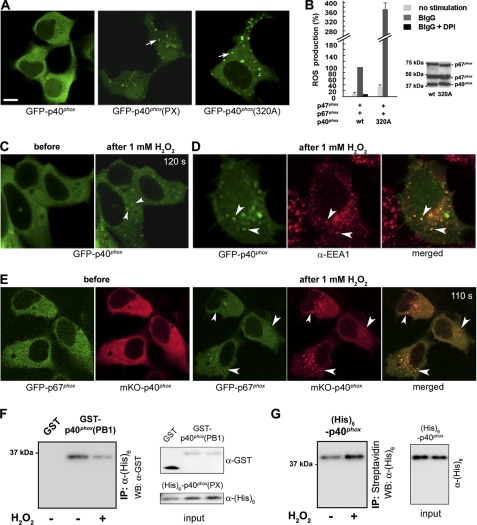
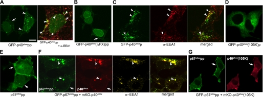
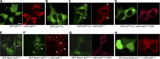
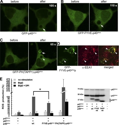
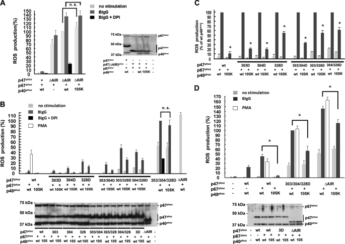
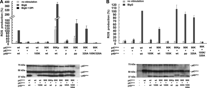
During activation of the phagocyte (Nox2-based) NADPH oxidase, the cytoplasmic Phox complex (p47(phox)-p67(phox)-p40(phox)) translocates and associates with the membrane-spanning flavocytochrome b(558). It is unclear where (in cytoplasm or on membranes), when (before or after assembly), and how p40(phox) acquires its PI(3)P-binding capabilities. We demonstrated that in addition to conformational changes induced by H(2)O(2) in the cytoplasm, p40(phox) acquires PI(3)P-binding through direct or indirect membrane targeting. We also found that p40(phox) is essential when p47(phox) is partially phosphorylated during FcγR-mediated oxidase activation; however, p40(phox) is less critical when p47(phox) is adequately phosphorylated, using phosphorylation-mimicking mutants in HEK293(Nox2/FcγRIIa) and RAW264.7(p40/p47KD) cells. Moreover, PI binding to p47(phox) is less important when the autoinhibitory PX-PB1 domain interaction in p40(phox) is disrupted or when p40(phox) is targeted to membranes. Furthermore, we suggest that high affinity PI(3)P binding of the p40(phox) PX domain is critical during its accumulation on phagosomes, even when masked by the PB1 domain in the resting state. Thus, in addition to mechanisms for directly acquiring PI(3)P binding in the cytoplasm by H(2)O(2), p40(phox) can acquire PI(3)P binding on targeted membranes in a p47(phox)-dependent manner and functions both as a "carrier" of the cytoplasmic Phox complex to phagosomes and an "adaptor" of oxidase assembly on phagosomes in cooperation with p47(phox), using positive feedback mechanisms.


Figures

FIGURE 1.
FIGURE 1.
H2O2 induces EE targeting of p40phox. A, transfected GFP-p40phox is localized in the cytoplasm (left); in contrast, GFP-p40phox(PX) (center; arrow) and p40phox(F320A) (right; arrow) are localized at dotlike structures, in resting WT HEK293 cells. Bar, 10 μm. B, p40phox(F320A) co-expressed with p47phox + p67phox supports moderate constitutive (32.0 ± 6.2%) and high BIgG-stimulated ROS production in HEK293Nox2/FcγRIIa cells when compared with cells expressing WT p40phox + p47phox + p67phox. Western blotting detects comparable levels of cytoplasmic Phox proteins in both transfection experiments. C, in response to exogenous 1 mm H2O2 (120 s after stimulation), GFP-p40phox expressed in WT HEK293 cells translocates to dotlike structures (arrowheads). D, the dotlike structures are co-stained by an EE marker, EEA1 (arrowheads). E, cytoplasmic GFP-p67phox co-expressed with mKO-p40phox in WT HEK293 cells translocates to dotlike structures (arrowheads) after stimulation (110 s) with 1 mm H2O2. Similar effects of H2O2 are observed in RAW 264.7 cells (supplemental Fig. 3). F, effect of H2O2 on the PX-PB1 domain interaction in vitro. The interaction between purified His6-p40phox(PX) and GST-p40phox(PB1) proteins detected by pull-down assays is weakened by the addition of 0.01 mm H2O2. Similar results are obtained in three independent experiments. G, effect of H2O2 on binding of p40phox to PI(3)P in vitro. The interaction between purified full-length His6-p40phox protein and PI(3)P-containing liposomes detected by pull-down assays is strengthened by the addition of 0.01 mm H2O2. Similar results were obtained in three independent experiments. Error bars, S.E.
FIGURE 2.
FIGURE 2.
Direct or indirect membrane targeting of p40phox induces EE-targeting of p40phox in WT HEK293 cells. A, transfected GFP-p40phoxpp is localized at dotlike structures (left; arrowheads) in addition to PM (left; arrow). The dotlike structures are co-stained with EEA1 (right; arrowheads). Bar, 10 μm. B, the dotlike localization of GFP-p40phoxpp disappears in the case of GFP-p40phox(ΔPX)pp. Arrow, PM. C, GFP-p40phoxp is co-stained with EEA1 (arrowheads). A movie of accumulation on phagosome of p40phoxp during ingestion of BIgG in HEK293Nox2/FcγRIIa cells is available (supplemental Video 1). D, the dotlike localization of GFP-p40phoxp disappears in the case of GFP-p40phox(R105K)p. E, PM-targeting mutant of cytoplasmic GFP-p67phox, GFP-p67phoxpp, shows PM localization (arrows) in addition to nuclear localization. F, when cytoplasmic mKO-p40phox is co-expressed with GFP-p67phoxpp, both proteins are co-localized at EEs stained by EEA1 (using Cy5-conjugated secondary Ab) (arrowheads) in addition to the PM (arrow). G, the dotlike localization of both proteins disappears in the case of mKO-p40phox(R105K) + GFP-p67phoxpp. Arrow, PM.
FIGURE 3.
FIGURE 3.
Indirect membrane targeting of p40phoxby p47phoxp or Noxo1-p47phox through the p47phox-p67phox-p40phox interaction induces EE localization of p40phox in HEK293p67phox cells. A, both GFP-p47phox and mKO-p40phox are localized in the cytoplasm. Bar, 10 μm. B, GFP-p47phoxp has a reticular, nuclear membrane and Golgi complex localization. C, in the case of co-expression of GFP-p47phoxp and mKO-p40phox, dotlike structures with GFP-p47phoxp and mKO-p40phox appear (arrows). Co-staining of stably expressed p67phox at the dotlike structures is shown in supplemental Fig. 5B. D, the dotlike structures disappear in the case of co-expression of GFP-p47phoxp and mKO-p40phox(R105K). E, GFP-Noxo1-p47phox shows PM localization (arrow) in addition to nuclear localization. F, with co-expression of GFP-Noxo1-p47phox and mKO-p40phox, mKO-p40phox is localized at dotlike structures (arrowheads) in addition to PM (arrow) with GFP-Noxo1-p47phox. In contrast, without co-expression of GFP-Noxo1-p47phox, mKO-p40phox is localized in the cytoplasm (asterisks). G, the dotlike localization, but not PM localization, of both proteins disappears in the case of co-expression of GFP-Noxo1-p47phox and mKO-p40phox(R105K). H, both the dotlike and PM localizations of both proteins disappear with co-expression of GFP-Noxo1(R40Q)-p47phox and mKO-p40phox.
FIGURE 4.
FIGURE 4.
High affinity PI(3)P-binding PX domain is a critical determinant of p40phox phagosomal accumulation and oxidase function in HEK293Nox2/FcγRIIa cells. A, weak accumulation of GFP-p40phox on phagosome 105 s after the start of BIgG ingestion is shown (arrow). A movie is available (supplemental Video 2). B, no accumulation of GFP-FYVE-p40phox on phagosome is observed (arrow). A movie is available (supplemental Video 3). C, accumulation of GFP-PH(TAPP1)-p40phox on nascent phagosomal cup is observed 65 s after starting BIgG ingestion (arrow). No accumulation of GFP-PH(TAPP1)-p40phox on mature phagosome is observed (arrowhead). A movie is available (supplemental Video 4). D, FYVE-p40phoxp is localized at dotlike structures that co-stained with EEA1 (arrowheads). A movie of accumulation on phagosome of FYVE-p40phoxp during ingestion of BIgG is available (supplemental Video 5). E, BIgG-stimulated ROS production is enhanced 4-fold by p47phox + p67phox + PH(TAPP1)-p40phox and is statistically decreased by p47phox + p67phox + FYVE-p40phox when compared with p47phox + p67phox + p40phox. *, p < 0.05. Error bars, S.E.
FIGURE 5.
FIGURE 5.
Dependence of PI(3)P-binding of p40phox in ROS production by Nox2 is determined by the phosphorylation status of p47phox in both HEK293Nox2/FcγRIIa (A–C) and RAW264.7p40/p47KD cells (D). A, ROS production using p47phox(ΔAIR) is constitutive, shows small BIgG dependence, and is effectively inhibited by 10 μm diphenylene iodonium. The addition of p40phox enhances ROS production by p47phox(ΔAIR) and p67phox. ROS production by p47phox(ΔAIR) + p67phox + p40phox is independent of the PI(3)P-binding capabilities of p40phox (p40phox(R105K)). n.s., not statistically significant. B, ROS production using one- or two-site phosphorylation-mimicking mutants of p47phox as well as WT p47phox shows PI(3)P-binding of p40phox dependence; however, p47phox(S303D/S304D/S328D) is independent of PI(3)P binding to p40phox. All mimicking mutants showed higher ROS production than WT p47phox. 3D, S303D/S304D/S328D. C, PI(3)P dependence in ROS production using WT or one- or two-site phosphorylation-mimicking mutants of p47phox. Data shown in B are normalized to activities supported by WT p40phox. All mimicking mutants show less PI(3)P dependence than WT p47phox. The mimicking mutants having Asp-328 (S328D, S303D/S328D, and S304D/S328D) show constitutive activity without BIgG and less PI(3)P dependence than S303D, S304D, or S303D/S304D. *, p < 0.01. D, in RAW264.7p40/p47KD cells, ROS production using WT p47phox shows almost complete dependence on PI(3)P binding to p40phox. In contrast, reconstituted ROS production by p47phox(S303D/S304D/S328D) or p47phox(ΔAIR) shows only moderate PI(3)P dependence. *, p < 0.01. 3D, S303D/S304D/S328D. Error bars, S.E.
FIGURE 6.
FIGURE 6.
Membrane-associating mutants of p40phox can rescue the function of the PX domain of p47phox. A, in HEK293Nox2/FcγRIIa cells, ROS production by p47phox(R90K) + p67phox + p40phox is almost absent, whereas p47phox(R90K)p dramatically rescues reconstituted ROS production. p40phoxpp, but not p40phoxp, is able to completely rescue ROS production by p47phox(R90K) + p67phox + p40phox. p40phox(F320A), which is targeted to EE by disrupted PX-PB1 intermolecular interaction (Fig. 1A), effectively rescues ROS production by p47phox(R90K) + p67phox + p40phox, whereas p40phox(R105K/F320A) does not. B, in RAW264.7p40/p47KD cells, WT p40phox, but not p40phox(R105K), moderately rescues ROS production with p47phox(R90K). Membrane-associating mutants, p47phox(R90K)p, p40phoxpp, and p40phox(F320A) almost completely rescue reconstituted ROS production with p47phox(R90K). Error bars, S.E.

Similar articles

Cited by

References

    1. Quinn M. T., Gauss K. A. (2004) J. Leukoc. Biol. 76, 760–781 - PubMed
    1. Sumimoto H. (2008) FEBS J. 275, 3249–3277 - PubMed
    1. Leto T. L., Morand S., Hurt D., Ueyama T. (2009) Antioxid. Redox Signal. 11, 2607–2619 - PMC - PubMed
    1. Borregaard N., Heiple J. M., Simons E. R., Clark R. A. (1983) J. Cell Biol. 97, 52–61 - PMC - PubMed
    1. Lapouge K., Smith S. J., Groemping Y., Rittinger K. (2002) J. Biol. Chem. 277, 10121–10128 - PubMed

Publication types

MeSH terms