

Primary cilia and coordination of receptor tyrosine kinase (RTK) signalling
- PMID: 21956154
- PMCID: PMC4294548
- DOI: 10.1002/path.3004
Primary cilia and coordination of receptor tyrosine kinase (RTK) signalling
Abstract
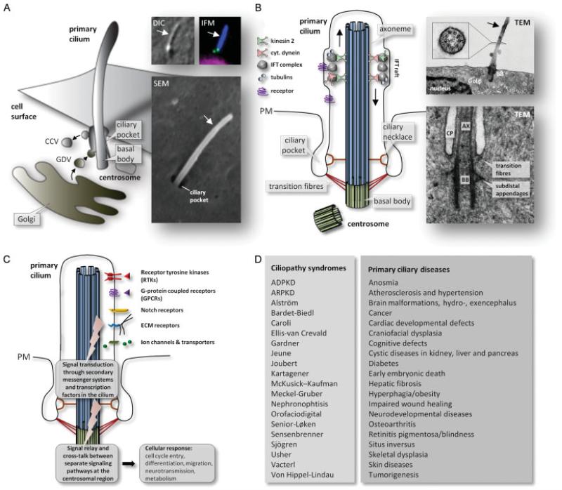
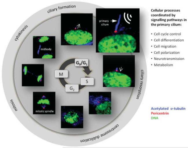
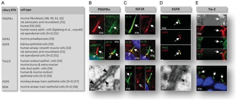
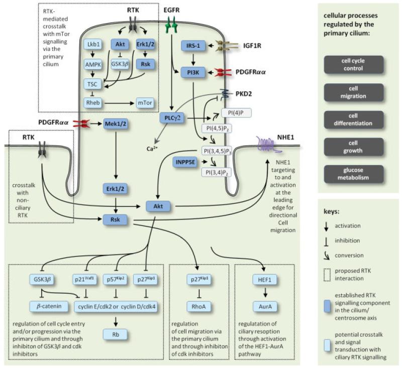
Primary cilia are microtubule-based sensory organelles that coordinate signalling pathways in cell-cycle control, migration, differentiation and other cellular processes critical during development and for tissue homeostasis. Accordingly, defects in assembly or function of primary cilia lead to a plethora of developmental disorders and pathological conditions now known as ciliopathies. In this review, we summarize the current status of the role of primary cilia in coordinating receptor tyrosine kinase (RTK) signalling pathways. Further, we present potential mechanisms of signalling crosstalk and networking in the primary cilium and discuss how defects in ciliary RTK signalling are linked to human diseases and disorders.
Copyright © 2011 Pathological Society of Great Britain and Ireland. Published by John Wiley & Sons, Ltd.
Figures
Similar articles
-
Primary Cilia and Coordination of Receptor Tyrosine Kinase (RTK) and Transforming Growth Factor β (TGF-β) Signaling.Cold Spring Harb Perspect Biol. 2017 Jun 1;9(6):a028167. doi: 10.1101/cshperspect.a028167. Cold Spring Harb Perspect Biol. 2017. PMID: 27638178 Free PMC article. Review.
-
The primary cilium coordinates signaling pathways in cell cycle control and migration during development and tissue repair.Curr Top Dev Biol. 2008;85:261-301. doi: 10.1016/S0070-2153(08)00810-7. Curr Top Dev Biol. 2008. PMID: 19147009 Review.
-
Receptor tyrosine kinase (RTK) signalling in the control of neural stem and progenitor cell (NSPC) development.Mol Neurobiol. 2014 Feb;49(1):440-71. doi: 10.1007/s12035-013-8532-5. Epub 2013 Aug 28. Mol Neurobiol. 2014. PMID: 23982746 Review.
-
Cellular signalling by primary cilia in development, organ function and disease.Nat Rev Nephrol. 2019 Apr;15(4):199-219. doi: 10.1038/s41581-019-0116-9. Nat Rev Nephrol. 2019. PMID: 30733609 Free PMC article. Review.
-
Emerging mechanistic understanding of cilia function in cellular signalling.Nat Rev Mol Cell Biol. 2024 Jul;25(7):555-573. doi: 10.1038/s41580-023-00698-5. Epub 2024 Feb 16. Nat Rev Mol Cell Biol. 2024. PMID: 38366037 Free PMC article. Review.
Cited by
-
Rab23 activities and human cancer-emerging connections and mechanisms.Tumour Biol. 2016 Oct;37(10):12959-12967. doi: 10.1007/s13277-016-5207-7. Epub 2016 Jul 23. Tumour Biol. 2016. PMID: 27449041 Review.
-
The Akt signaling pathway is required for tissue maintenance and regeneration in planarians.BMC Dev Biol. 2016 Apr 11;16:7. doi: 10.1186/s12861-016-0107-z. BMC Dev Biol. 2016. PMID: 27068018 Free PMC article.
-
The essential role of primary cilia in cerebral cortical development and disorders.Curr Top Dev Biol. 2021;142:99-146. doi: 10.1016/bs.ctdb.2020.11.003. Epub 2021 Jan 25. Curr Top Dev Biol. 2021. PMID: 33706927 Free PMC article. Review.
-
Reciprocal Regulation between Primary Cilia and mTORC1.Genes (Basel). 2020 Jun 26;11(6):711. doi: 10.3390/genes11060711. Genes (Basel). 2020. PMID: 32604881 Free PMC article. Review.
-
Signaling through the Primary Cilium.Front Cell Dev Biol. 2018 Feb 8;6:8. doi: 10.3389/fcell.2018.00008. eCollection 2018. Front Cell Dev Biol. 2018. PMID: 29473038 Free PMC article. Review.
References
-
- Badano JL, Mitsuma N, Beales PL, et al. The ciliopathies: an emerging class of human genetic disorders. Annu Rev Genom Hum Genet. 2006;7:125–148. - PubMed
-
- Fliegauf M, Benzing T, Omran H. When cilia go bad: cilia defects and ciliopathies. Nat Rev Mol Cell Biol. 2007;8:880–893. - PubMed
-
- Christensen ST, Pedersen LB, Schneider L, et al. Sensory cilia and integration of signal transduction in human health and disease. Traffic. 2007;8:97–109. - PubMed
Publication types
MeSH terms
Substances
Grants and funding
LinkOut - more resources
Full Text Sources
