. 2012 Mar;8(1):71-80.
doi: 10.1007/s11302-011-9262-7. Epub 2011 Sep 29.

Purine receptors and Ca(2+) signalling in the human blood-brain barrier endothelial cell line hCMEC/D3

Affiliations

Purine receptors and Ca(2+) signalling in the human blood-brain barrier endothelial cell line hCMEC/D3

Willem Bintig et al. Purinergic Signal. 2012 Mar.

Abstract

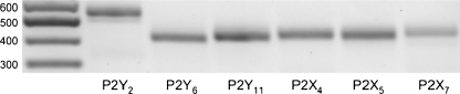
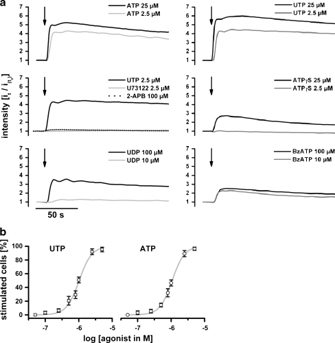
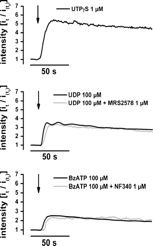
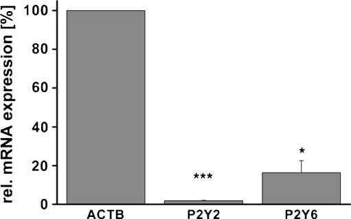
The expression and physiology of purine receptors of the human blood-brain barrier endothelial cells were characterised by application of molecular biological, gene-silencing and Ca(2+)-imaging techniques to hCMEC/D3 cells. Reverse transcription polymerase chain reaction showed the expression of the G-protein-coupled receptors P2Y(2)-, P2Y(6)-, P2Y(11)- as well as the ionotropic P2X(4)-, P2X(5)- and P2X(7)-receptors. Fura-2 ratiometry revealed that adenosine triphosphate (ATP) or uridine triphosphate (UTP) mediated a change in the intracellular Ca(2+) concentration ([Ca(2+)](i)) from 150 to 300 nM in single cells. The change in [Ca(2+)](i) corresponded to a fourfold to fivefold increase in the fluorescence intensity of Fluo-4, which was used for high-throughput experiments. Pharmacological dissection using different agonists [UTPγS, ATPγS, uridine diphosphate (UDP), adenosine diphosphate (ADP), BzATP, αβ-meATP] and antagonist (MRS2578 or NF340) as well as inhibitors of intracellular mediators (U73122 and 2-APB) showed a PLC-IP(3) cascade-mediated Ca(2+) release, indicating that the nucleotide-induced Ca(2+) signal was mainly related to P2Y(2, 6 and 11) receptors. The gene silencing of the P2Y(2) receptor reduced the ATP- or UTP-induced Ca(2+) signal and suppressed the Ca(2+) signal mediated by P2Y(6) and P2Y(11) more specific agonists like UDP (P2Y(6)), BzATP (P2Y(11)) and ATPγS (P2Y(11)). This report identifies the P2Y(2) receptor subtype as the main purine receptor involved in Ca(2+) signalling of the hCMEC/D3 cells.


Figures

Fig. 1
Fig. 1
The different purine receptors expressed by the hCMEC-D3 cells as revealed by the RT-PCR. Expression of P2Y2, 6 and 11 as well as P2X4, 5 and 7 subtypes by hCMEC/D3 cells was confirmed by RT-PCR. The original band for P2Y6 was almost undetectable. Another round of PCR was therefore performed following extraction of the original PCR band. The product of this re-PCR is visualised by agarose gel electrophoresis together with the original PCR products for the other purine receptor subtypes. The other P2X and P2Y receptor subtypes were not found
Fig. 2
Fig. 2
a Change in fluorescence intensity induced by stimulation of the cells with different agonist concentration. The data show that the cell sensitivity to ATP and UTP was similar. ATPγS, UDP or BzATP could also stimulate the cells. However, the change in fluorescence intensity was reduced (even at high concentrations) in comparison to stimulation with ATP or UTP. The PLC inhibitor U73122 or the IP3 receptor blocker 2-ABP could block the UTP or ATP induced increase in fluorescence intensity. The graphs represent averages of at least 200 cells for each treatment. For the sake of clarity, the error bars are not shown. The maximal SEM was ±0.21. b The concentration dependency of stimulation of the Ca2+ signal in the hCMEC/D3 cells by the putative physiological agonists. The portion of cells stimulated by respective ATP or UTP concentrations is given. The data points were fitted to sigmoid dose–response curves. This allowed to estimate a half stimulation concentration of about 1.25 μM for both agonists
Fig. 3
Fig. 3
The specific agonist of P2Y2 and P2Y4 receptor UTPγS induced Ca2+ signals in the hCMEC/D3 cells. Since the cells did not express P2Y4 receptor (Fig. 1) the signal is related to P2Y2 receptors. Additionally, MRS2578 or NF340 the specific inhibitors of P2Y6 and P2Y11 respectively did not affect the UDP or the BzATP induced Ca2+ signal in hCMEC/D3 cells
Fig. 4
Fig. 4
The gene silencing of P2Y2 and P2Y6 receptor using specific siRNAs was estimated by quantitative RT-PCR experiments. Relative to negative siRNA, the P2Y2 siRNA (s9967) and the P2Y6 siRNA (s224151) reduced the mRNA for the receptors by 98% and 83.7%, respectively. The data were normalised to the housekeeping gene hACTB
Fig. 5
Fig. 5
Silencing the P2Y2 receptor reduced the sensitivity of the cells to UTP and suppressed the sensitivity to UDP, ATPγS, BzATP. Cells in which the P2Y6 was silenced by specific siRNA still react to UDP stimulation. It is noteworthy that the negative siRNA reduced the sensitivity of the cells to the agonists. This is probable due to a nonspecific effect related to the Lipofectamine 2000 used during the transfection

References

    1. Drewes LR. Molecular architecture of the brain microvasculature: perspective on blood–brain transport. J Mol Neurosci. 2001;16:93–98. doi: 10.1385/JMN:16:2-3:93. - DOI - PubMed
    1. Abbott NJ, Rönnbäck L, Hansson E. Astrocyte–endothelial interactions at the blood–brain barrier. Nat Rev Neurosci. 2006;7:41–53. doi: 10.1038/nrn1824. - DOI - PubMed
    1. Banks WA. Physiology and pathology of the blood–brain barrier: implications for microbial pathogenesis, drug delivery and neurodegenerative disorders. J Neurovirol. 1999;6:538–555. doi: 10.3109/13550289909021284. - DOI - PubMed
    1. Iadecola C, Nedergaard M. Glial regulation of the cerebral microvasculature. Nat Neurosci. 2007;10:1369–1376. doi: 10.1038/nn2003. - DOI - PubMed
    1. Zoppo GJ. Virchow’s triad: the vascular basis of cerebral injury. Rev Neurol Dis. 2008;5:S12–S21. - PMC - PubMed

LinkOut - more resources