. 2012 Jan;37(2):499-507.
doi: 10.1038/npp.2011.215. Epub 2011 Sep 28.

Investigation of anatomical thalamo-cortical connectivity and FMRI activation in schizophrenia

Affiliations

Investigation of anatomical thalamo-cortical connectivity and FMRI activation in schizophrenia

Stefano Marenco et al. Neuropsychopharmacology. 2012 Jan.

Abstract

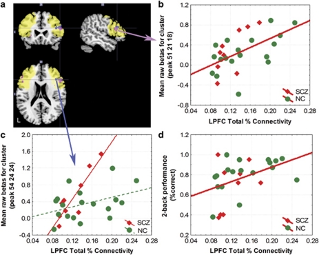
The purpose of this study was to examine measures of anatomical connectivity between the thalamus and lateral prefrontal cortex (LPFC) in schizophrenia and to assess their functional implications. We measured thalamocortical connectivity with diffusion tensor imaging (DTI) and probabilistic tractography in 15 patients with schizophrenia and 22 age- and sex-matched controls. The relationship between thalamocortical connectivity and prefrontal cortical blood-oxygenation-level-dependent (BOLD) functional activity as well as behavioral performance during working memory was examined in a subsample of 9 patients and 18 controls. Compared with controls, schizophrenia patients showed reduced total connectivity of the thalamus to only one of six cortical regions, the LPFC. The size of the thalamic region with at least 25% of model fibers reaching the LPFC was also reduced in patients compared with controls. The total thalamocortical connectivity to the LPFC predicted working memory task performance and also correlated with LPFC BOLD activation. Notably, the correlation with BOLD activation was accentuated in patients as compared with controls in the ventral LPFC. These results suggest that thalamocortical connectivity to the LPFC is altered in schizophrenia with functional consequences on working memory processing in LPFC.


Figures

Figure 1
Figure 1
Cortical ROIs in one healthy volunteer. The subdivisions of the cortex used in the analysis are shown for the left hemisphere in a lateral (top) and medial (bottom) view with a color bar for reference. Bilateral ROIs were used in the analysis, whereas unilateral ROIs are shown here for visualization. The following original Freesurfer parcellations (Desikan et al, 2006) were combined to form the larger ROIs used here: orbitofrontal cortex (OFC: pars orbitalis, medial orbitofrontal cortex, lateral orbitofrontal cortex), medial prefrontal cortex (MPFC: caudal anterior cingulate, rostral anterior cingulate, superior frontal gyrus), lateral prefrontal cortex (LPFC: pars triangularis, frontal pole, rostral middle frontal gyrus, pars opercularis), sensorimotor cortex (SMC: precentral gyrus, caudal middle frontal gyrus, post-central gyrus, paracentral lobule), parietal cortex (PC: inferior parietal cortex, supramarginal gyrus, precuneus cortex, posterior cingulate cortex, isthmus cingulate, superior parietal cortex), medial temporal cortex (MTC: entorhinal cortex, parahippocampal gyrus, fusiform gyrus), lateral temporal cortex (LTC: transverse temporal cortex, superior temporal gyrus, banks of the superior temporal sulcus, inferior temporal gyrus, middle temporal gyrus, temporal pole), and occipital cortex (OCC: pericalcarine cortex, lingual gyrus, lateral occipital cortex, cuneus cortex). Graphics were created using SUMA, part of the AFNI package (http://afni.nimh.nih.gov/afni/suma).
Figure 2
Figure 2
Results for total percent connectivity and CDR size. (a) Total percent connectivity for the six cortical ROIs included in the analysis. (b) Thalamic CDR size for the six regions included in the analysis. P values represent post-hoc unpaired t-tests. *P<0.05 on the post-hoc tests. Error bars represent SD. LTC, lateral temporal cortex; LPFC, lateral PFC; MPFC, medial prefrontal cortex; OCC, occipital cortex; PC, parietal cortex; SMC, somato-motor cortex.
Figure 3
Figure 3
Comparison of the spatial consistency of LPFC thalamic CDRs between patients with schizophrenia and controls. Binary masks of the thalamic CDRs for the healthy volunteers (n=22) and patients with schizophrenia (n=15) were normalized to a standard template (Wake Forest University Pickatlas: http://fmri.wfubmc.edu/cms/softwarePickAtlas), averaged, and thresholded to show only those voxels with >10% of the subjects represented at that location. The black border outlines five slices of the thalamus, oriented from top to bottom, with the axes showing the mm coordinates in standard MNI space. Warmer colors illustrate a higher spatial consistency across subjects, with dark red representing 100% of subjects present at a particular location. Graphics were created using Matlab (Mathworks, Natick, MA).
Figure 4
Figure 4
Correlation of prefrontal BOLD activation and LPFC total percent connectivity. (a) The significant clusters are shown superimposed on the MNI single subject template. The yellow area represents the region of interest used to constrain the analysis, the pink area corresponds to the cluster (maximum at voxel coordinates 51 21 18) where both groups had similar positive slopes for the correlation between LPFC total percent connectivity and BOLD, whereas the blue area corresponds to the cluster (maximum at voxel 54 24 24) where slopes differed significantly across groups. (b) Correlation between LPFC total percent connectivity and BOLD parameter estimates for the cluster with the peak at voxel 51 21 18 (raw β values adjusted for the session mean of the individual subject, n=27, r=0.56, p=0.0027, z for peak voxel=3.33). The cluster surrounding the peak of significance encompassed 24 voxels (648 μl). This exceeded the threshold of 23 voxels (621 μl) found by AlphaSim and can therefore be considered corrected for multiple comparisons. (c) Significant difference in the correlation between LPFC connectivity and BOLD activation in the cluster surrounding voxel 54 24 24 (raw β values adjusted for the session mean, normal controls (NC): n=18, r=0.4, patients with schizophrenia (SCZ): n=9, r=0.9, 24 voxel extent or 648 μl, z for peak voxel=3.38, p=0.0007, uncorrected). (d) Correlation between LPFC connectivity and performance on the 2-back working memory task (r=0.43, p=0.02) for the whole group. Correlation coefficients for patients and controls did not differ from each other (SCZ=0.38, NOR=0.44, Fischer's transformation: z=−0.15, p=0.88).

References

    1. Adriano F, Spoletini I, Caltagirone C, Spalletta G. Updated meta-analyses reveal thalamus volume reduction in patients with first-episode and chronic schizophrenia. Schizophr Res. 2011;123:1–14. - PubMed
    1. Agarwal N, Rambaldelli G, Perlini C, Dusi N, Kitis O, Bellani M, et al. Microstructural thalamic changes in schizophrenia: a combined anatomic and diffusion weighted magnetic resonance imaging study. J Psychiatry Neurosci. 2008;33:440–448. - PMC - PubMed
    1. Akbarian S, Vinuela A, Kim JJ, Potkin SG, Bunney WE, Jr, Jones EG. Distorted distribution of nicotinamide-adenine dinucleotide phosphate-diaphorase neurons in temporal lobe of schizophrenics implies anomalous cortical development. Arch Gen Psychiatry. 1993;50:178–187. - PubMed
    1. Anderson SA, Volk DW, Lewis DA. Increased density of microtubule associated protein 2-immunoreactive neurons in the prefrontal white matter of schizophrenic subjects. Schizophr Res. 1996;19:111–119. - PubMed
    1. Andreasen NC, O'Leary DS, Cizadlo T, Arndt S, Rezai K, Ponto LL, et al. Schizophrenia and cognitive dysmetria: a positron-emission tomography study of dysfunctional prefrontal-thalamic-cerebellar circuitry. Proc Natl Acad Sci USA. 1996;93:9985–9990. - PMC - PubMed

Publication types

MeSH terms