Review
. 2010 Jul-Aug;1(1):47-59.
doi: 10.1002/wrna.3. Epub 2010 May 6.

Viruses and the cellular RNA decay machinery

Affiliations
Review

Viruses and the cellular RNA decay machinery

Marta Maria Gaglia et al. Wiley Interdiscip Rev RNA. 2010 Jul-Aug.

Abstract

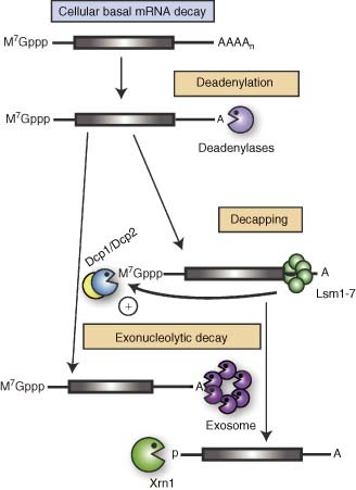
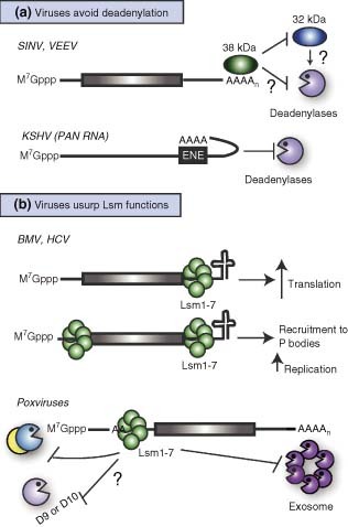
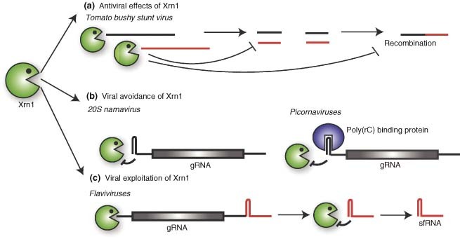
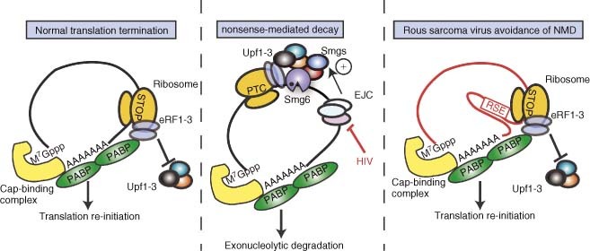
The ability to control cellular and viral gene expression, either globally or selectively, is central to a successful viral infection, and it is also crucial for the host to respond and eradicate pathogens. In eukaryotes, regulation of message stability contributes significantly to the control of gene expression and plays a prominent role in the normal physiology of a cell as well as in its response to environmental and pathogenic stresses. Not surprisingly, emerging evidence indicates that there are significant interactions between the eukaryotic RNA turnover machinery and a wide variety of viruses. Interestingly, in many cases viruses have evolved mechanisms not only to evade eradication by these pathways, but also to manipulate them for enhanced viral replication and gene expression. Given our incomplete understanding of how many of these pathways are normally regulated, viruses should be powerful tools to help deconstruct the complex networks and events governing eukaryotic RNA stability.


Figures

Figure 1
Figure 1
Basal pathways of cellular mRNA decay. Degradation of normal cellular mRNA initiates with removal of the poly(A) tail by one of the cellular deadenylases. Subsequently, the Lsm1–7 protein complex binds the 3 untranslated region (UTR) of the deadenylated messages and stimulates decapping by the Dcp2 enzyme and its activator protein Dcp1. The message body is subject to 3′→5′ exonucleolytic decay by the exosome or 5′→3′ decay by Xrn1.
Figure 2
Figure 2
Viral circumvention or utilization of cellular deadenylation and decapping pathways. (a) The RNAs of Sindbis virus (SINV) and Venezuelan equine encephalitis virus (VEEV) contains elements in the 3 UTR that recruit a cellular 38‐kDa protein and block deadenylation, presumably by preventing binding of a 32‐kDa deadenylation‐promoting factor. The PAN RNA of Kaposi's sarcoma‐associated herpesvirus contains an element (ENE) that interacts in cis with the poly(A) tail thus preventing deadenylase access. (b) The RNA genomes of Brome mosaic virus (BMV) and hepatitis C virus (HCV) can be bound by Lsm proteins in their 3 and/or 5 UTRs. This enhances their translation, as well as facilitates replication, possibly through recruitment of the RNAs to P bodies. Poxviruses transcripts have polyadenine sequences within their 5 UTRs, which bind Lsm1–7. Lsm binding prevents decapping (and presumably 5′→3′ decay), as well as 3′→5′ degradation of the messages.
Figure 3
Figure 3
Viral interactions with Xrn1. (a) During infection with Tomato bushy stunt virus, Xrn1 can prevent the accumulation of viral RNA and of viral RNA intermediates that serve as substrates for recombination. (b) Some viruses prevent Xrn1 degradation of their uncapped genomic RNAs (gRNA) by having highly structured sequences in the 5 UTR, as in the case of 20S narnavirus, or by recruiting protein complexes to this region, as in the case of Picornaviruses. (c) Flaviviral RNAs contain Xrn1 blocking sequences in their 3 UTR. Xrn1‐mediated degradation of the RNAs up to the blocking sequence results in the generation of a functional subgenomic RNA, sfRNA.
Figure 4
Figure 4
Nonsense‐mediated mRNA decay and mechanisms of viral avoidance. (a) When the translation machinery reaches the stop codon of a normal mRNA, the eukaryotic release factors eRF1 and eRF3 are recruited to the ribosome, in part via interactions with PABP. This results in efficient translation termination and re‐initiation. (b) When mRNAs containing a premature stop codon (PTC) are translated, the presence of EJCs downstream of the PTC, coupled with inefficient interactions between PABP and the termination factors, triggers nonsense‐mediated decay (NMD). NMD is induced by recruitment of Upf1–3 and the Smg proteins; cleavage by the endonuclease Smg6 triggers degradation of the mRNA fragments. Viruses such as HIV‐1 regulate the alterative splicing of their genomic RNAs to avoid the presence of EJCs downstream of stop codons. (c) The unspliced RNA of Rous sarcoma virus has a very long 3 UTR, which is stabilized by the RSE element that directly inhibits NMD. This element may artificially ‘shorten’ the 3 UTR by making contacts with sequences close to the poly(A) tail.
Figure 5
Figure 5
No‐go decay (NGD) as an antiviral pathway. In yeast ectopically expressing the plant protein PAP, the RNAs of Brome mosaic virus are depurinated. This causes translating ribosomes to stall, thereby triggering NGD via recruitment of the endonuclease Dom34p to the translation complex. After Dom34p cleaves the RNA close to the site of stalling, the resulting fragments become targets for exonucleases such as Xrn1 and the exosome.
Figure 6
Figure 6
Viruses interfere with several stages of the RNase L pathway. RNase L is indirectly activated by the presence of double‐stranded RNA (dsRNA) species in the cytoplasm of infected cells. dsRNA activates 2,5‐oligoadenylate synthetase (OAS), which produces 2‐5‐oligoadenylate (2–5A), an allosteric activator for RNase L (asterisks indicate active enzymes). The NS1 protein of influenza virus A prevents OAS activation, whereas vaccinia virus and HIV‐1 block activation of RNase L by 2–5A, the latter by decreasing the affinity of RNase L for its activator. Once activated, RNase L can cleave viral RNAs and also cellular mRNAs and rRNAs. Several viruses specifically protect their RNAs from degradation, including poliovirus (PV), hepatitis C virus (HCV), and possibly reoviruses, using their σ 3 protein. Reoviruses also exploit RNase L‐mediated destruction of cellular messages to decrease competition for gene expression machinery.

References

    1. Doma MK, Parker R. RNA quality control in eukaryotes. Cell 2007, 131: 660–668. - PubMed
    1. Goldstrohm AC, Wickens M. Multifunctional deadenylase complexes diversify mRNA control. Nat Rev Mol Cell Biol 2008, 9: 337–344. - PubMed
    1. Parker R, Song H. The enzymes and control of eukaryotic mRNA turnover. Nat Struct Mol Biol 2004, 11: 121–127. - PubMed
    1. Garneau NL, Sokoloski KJ Opyrchal M, Neff CP, Wilusz CJ, et al. The 3 untranslated region of sindbis virus represses deadenylation of viral transcripts in mosquito and Mammalian cells. J Virol 2008, 82: 880–892. - PMC - PubMed
    1. Conrad NK, Steitz JA. A Kaposi's sarcoma virus RNA element that increases the nuclear abundance of intronless transcripts. Embo J 2005, 24: 1831–1841. - PMC - PubMed

Publication types