Review
. 2011 Mar-Apr;2(2):167-83.
doi: 10.1002/wrna.40. Epub 2010 Sep 15.

Mechanisms of deadenylation-dependent decay

Affiliations
Review

Mechanisms of deadenylation-dependent decay

Chyi-Ying A Chen et al. Wiley Interdiscip Rev RNA. 2011 Mar-Apr.

Abstract

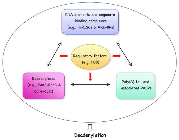
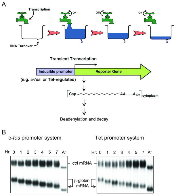
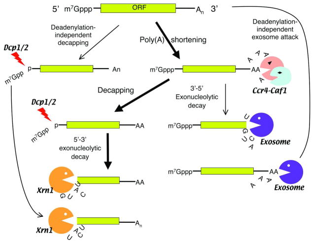
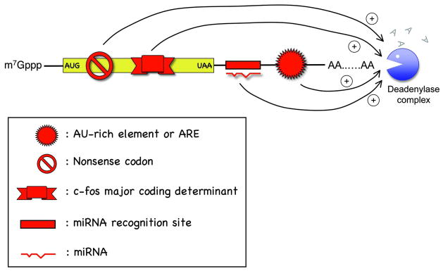
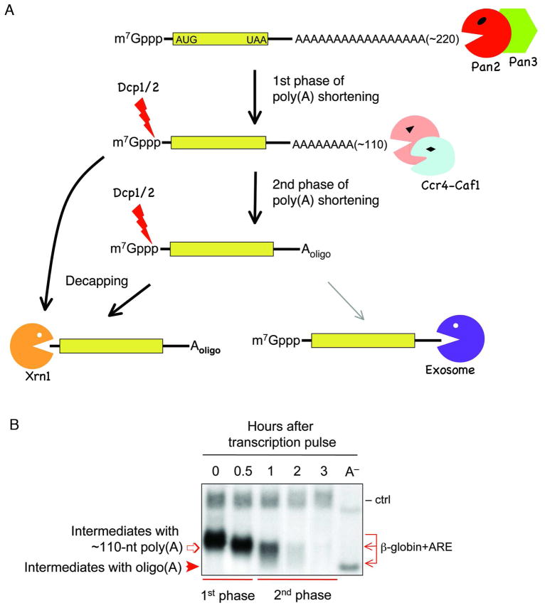
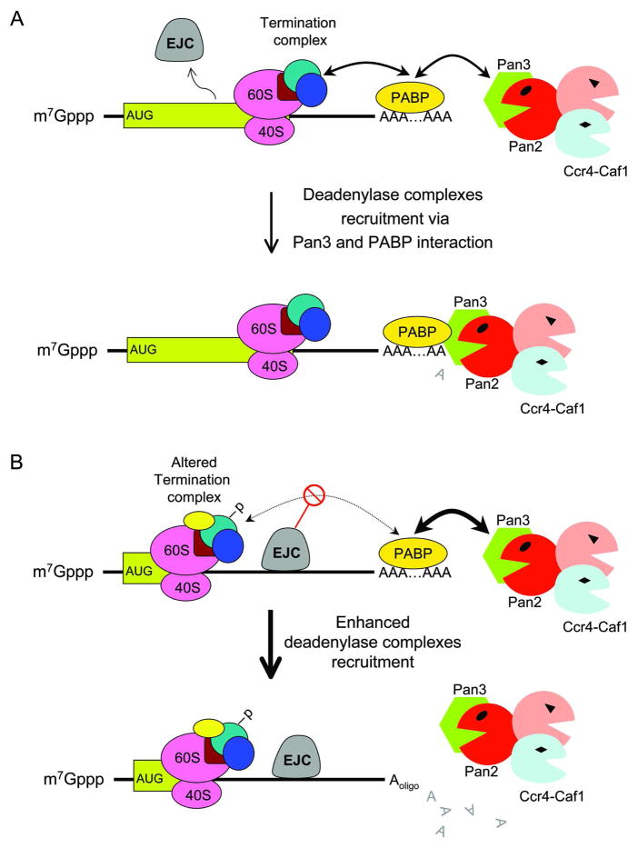
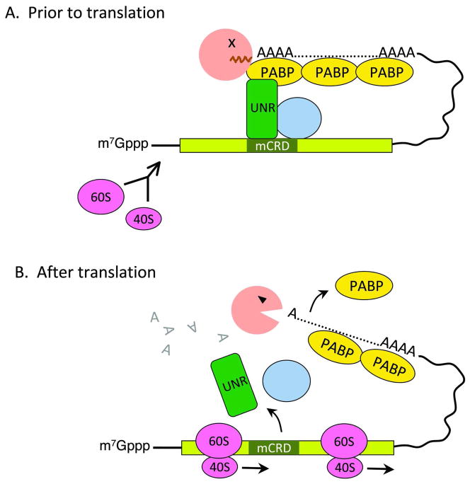
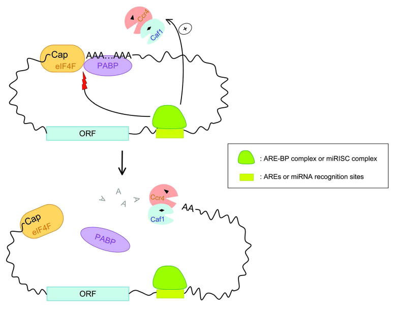
Degradation of messenger RNAs (mRNAs) plays an essential role in modulation of gene expression and in quality control of mRNA biogenesis. Nearly all major mRNA decay pathways characterized thus far in eukaryotes are initiated by deadenylation, i.e., shortening of the mRNA 3(') poly(A) tail. Deadenylation is often a rate-limiting step for mRNA degradation and translational silencing, making it an important control point for both processes. In this review, we discuss the fundamental principles that govern mRNA deadenylation in eukaryotes. We use several major mRNA decay pathways in mammalian cells to illustrate mechanisms and regulation of deadenylation-dependent mRNA decay, including decay directed by adenine/uridine-rich elements (AREs) in the 3(') -untranslated region (UTR), the rapid decay mediated by destabilizing elements in protein-coding regions, the surveillance mechanism that detects and degrades nonsense-containing mRNA [i.e., nonsense-mediated decay (NMD)], the decay directed by miRNAs, and the default decay pathway for stable messages. Mammalian mRNA deadenylation involves two consecutive phases mediated by the PAN2-PAN3 and the CCR4-CAF1 complexes, respectively. Decapping takes place after deadenylation and may serve as a backup mechanism to trigger mRNA decay if initial deadenylation is compromised. In addition, we discuss how deadenylation impacts the dynamics of RNA processing bodies (P-bodies), where nontranslatable mRNAs can be degraded or stored. Possible models for mechanisms of various deadenylation-dependent mRNA decay pathways are also discussed.


Figures

Figure 1
Figure 1
The deadenylation rate of an mRNA in mammalian cells is determined by interplays among several key players.
Figure 2
Figure 2
The transcriptional pulsing approach to determine mRNA deadenylation and decay kinetics. A. Schematic diagram illustrating how an inducible promoter can be used to achieve a short burst of mRNA production for kinetic studies. B. Northern blots showing default deadenylation exhibited by the stable β-globin mRNA. Transient transcription is driven by either the c-fos promoter (left) or the Tet-promoter (right). Times indicate hours (Hr) after transcriptional pulsing of β-globin gene. A : poly(A) mRNA as a size marker; ctrl : control mRNA constitutively expressed used as an internal control.
Figure 3
Figure 3
Schematic diagram showing eukarytoic mRNA decay pathways in the cytoplasm. Thick arrows indicate the major pathway for cytoplasmic mRNA degradation triggered by deadenylation. Note that the decay pathway triggered by an endonucleolytic cleavage (e.g., siRNA-mediated decay) is not depicted here.
Figure 4
Figure 4
Schematic diagram illustrating cis-acting elements or mutations in mammalian mRNA that promote accelerated deadenylation.
Figure 5
Figure 5
A: Schematic diagram showing the consecutive phases of deadenylation and mechanistic steps following deadenylation in mammalian cells. B: Northern blot showing that biphasic deadenylation precedes decay of the RNA body. Transient transcription of β-globin carrying the c-fos ARE is driven by the Tet-promoter. Times indicate hours after transcriptional pulsing of the gene. A : poly(A) mRNA as a size marker; ctrl : control mRNA constitutively expressed used as an internal control.
Figure 6
Figure 6
Schematic diagram showing a model for default deadenylation (A) and for acclerated deadenylation by nonsense codon (B) in mammalian cells. -P: modification of termination complex, e.g., phosphorylation. EJC: exon-exon junction complex.
Figure 7
Figure 7
A model for translationally coupled deadenylation mediated by the c-fos major coding region determinant of instability (mCRD). Prior to translation (A), Unr, other auxiliary factors (shown here as a golden yellow circle), and Ccr4 form a bridging complex that brings the 3′ poly(A) tail and the mCRD together. Ccr4 access to the poly(A) tail is blocked. Following translation initiation (B), ribosome transit knocks off the bridging complex and somehow activates Ccr4 for accelerated deadenylation.
Figure 8
Figure 8
Schematic diagram showing possible modes of accelerated deadenylation promoted by AREs or miRNA binding sites in mammalian cells, which involve either direct recruitment of Ccr4-Caf1 and/or disruption of eIF4F-PABP interaction to open up the closed loop formed between the 5′ Cap and 3′ poly(A) tail. Note that these two mechanisms are not mutually exclusive.

References

    1. Wickens M, et al. Translational control in developmental decisions. In: Mathews M, editor. Translational Control. 2. Cold Spring Harbor Press; 2000.
    1. Gebauer F, Hentze MW. Molecular Mechanisms of translational control. Nat Rev Mol Cell Biol. 2004;5:827–835. - PMC - PubMed
    1. Moore MJ. From birth to death: the complex lives of eukaryotic mRNAs. Science. 2005;309:1514–1518. - PubMed
    1. Garneau NL, et al. The highways and byways of mRNA decay. Nat Rev Mol Cell Biol. 2007;8:113–126. - PubMed
    1. Besse F, Ephrussi A. Translational control of localized mRNAs: restricting protein synthesis in space and time. Nat Rev Mol Cell Biol. 2008;9:971–980. - PubMed

Publication types

LinkOut - more resources