Review
. 2011 Mar-Apr;2(2):299-311.
doi: 10.1002/wrna.63. Epub 2011 Jan 12.

Recognition of S-adenosylmethionine by riboswitches

Affiliations
Review

Recognition of S-adenosylmethionine by riboswitches

Robert T Batey. Wiley Interdiscip Rev RNA. 2011 Mar-Apr.

Abstract

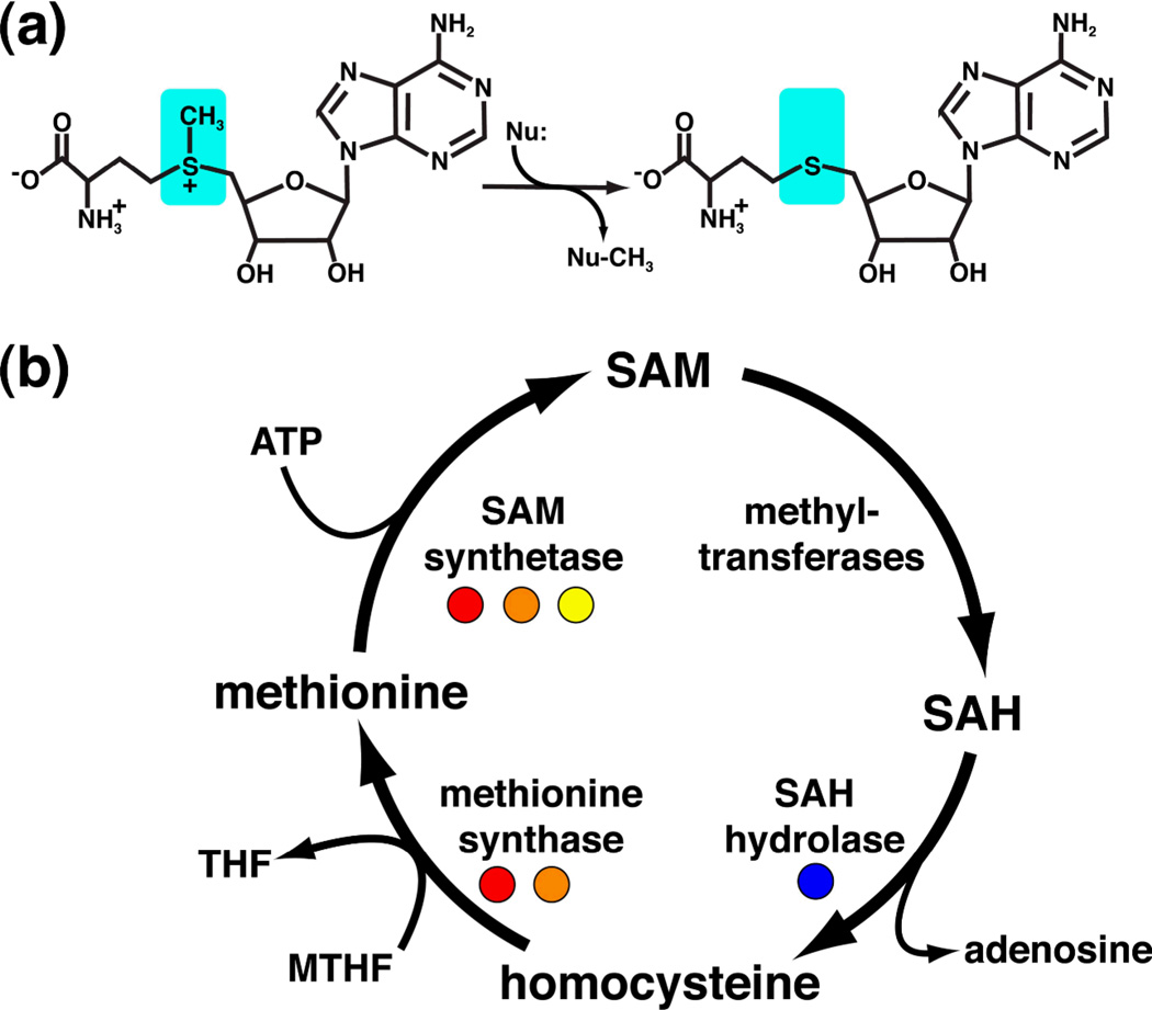
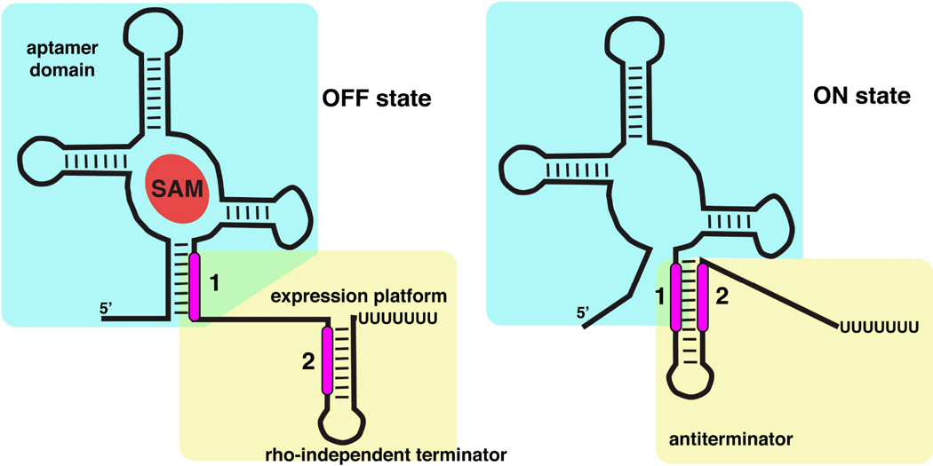
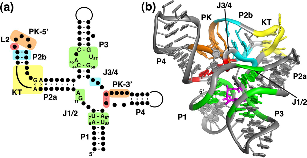
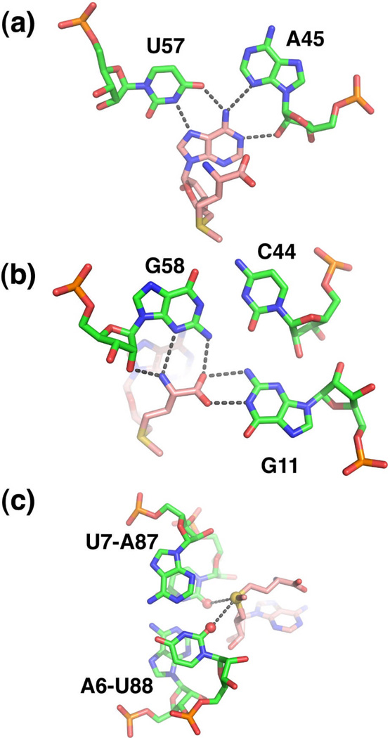
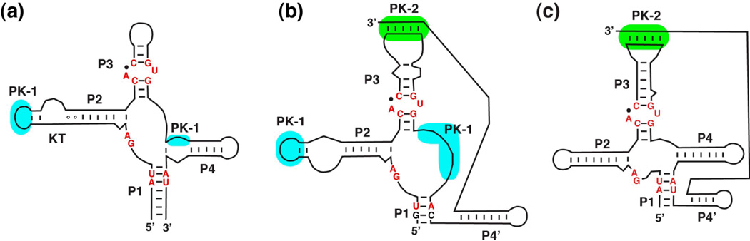
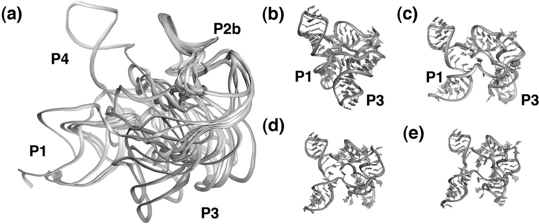
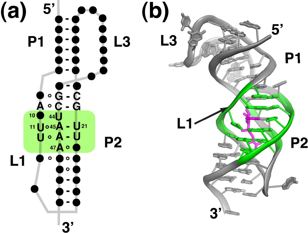
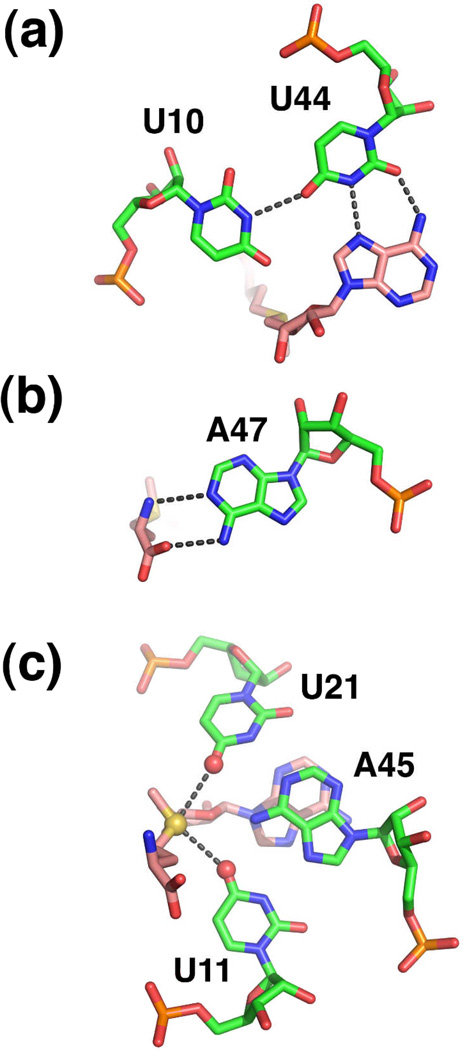
Riboswitches are regulatory elements commonly found in the 5' leader sequences of bacterial mRNAs that bind cellular metabolites to direct expression at either the transcriptional or translational level. The effectors of these RNAs are chemically diverse, including nucleobases and nucleosides, amino acids, cofactors, and second messenger molecules. Over the last few years, a number of structures have revealed the architectural means by which RNA creates binding pockets of high affinity and specificity for these compounds. For most effectors, there is a single class of associated riboswitches. However, eight individual classes of S-adenosylmethionine (SAM) and/or S-adenosylhomocysteine (SAH) responsive riboswitches that control various aspects of sulfur metabolism have been validated, revealing a diverse set of solutions to the recognition of these ubiquitous metabolites. This review focuses upon the structures of RNAs that bind SAM and SAH and how they discriminate between these compounds.


Figures

Figure 1
Figure 1
(a) Chemical structures of S-adenosyl-L-methionine (reactant) and S-adenosyl-L-homocysteine (product). SAM is the primary donor of methyl groups in biology via a nucleophilic attack (Nu) on its activated methyl group (cyan), such that SAH is the leaving group. (b) The SAM regeneration cycle. Enzymes regulated by SAM and SAH riboswitches are highlighted with colored circles (red, SAM-I; orange, SAM-II; yellow, SAM-III; blue, SAH).
Figure 2
Figure 2
Alternative secondary structures of a riboswitch that regulates transcription. The "off" state (left) is characterized by SAM bound to the aptamer domain (cyan box) which incorporates a sequence that is shared with the downstream expression platform (marked as "1"). This forces the expression platform (yellow box) to form a terminator stem-loop. In the "on" state (right) the lack of SAM binding allows the expression platform to form an antiterminator element using the sequence shared between the two domains. Note the mutual exclusivity of formation of the antiterminator and terminator through differential partitioning of sequences "1" and "2".
Figure 3
Figure 3
Structure of the SAM-I riboswitch. (a) Phylogenetically conserved primary and secondary structure of the class I SAM riboswitch. Nucleotides that are >97% conserved are specified by their identity; conserved secondary structure is shown as dots. Colored boxes refer to elements of conserved tertiary architecture: a kink-turn motif (yellow), a pseudoknot (orange), adenosine minor triples (cyan), and a base triple (red) as well as structure directly involved in SAM binding (green). Note that the all of the conserved residues are either in the critical G-A pairs that define the kink-turn motif or in the SAM binding pocket. The numbering of key nucleotides is consistent with that in Montange and Batey. (b) Cartoon representation of the X-ray crystal structure of the SAM-I riboswitch aptamer domain bound to SAM. This panel and other molecular representations of the SAM-I riboswitch were made using the coordinates in PDB accession number 3GX5. The color scheme is the same as in section (a), emphasizing the placement of the conserved tertiary architecture and the SAM binding pocket. SAM is shown as magenta sticks.
Figure 4
Figure 4
Recognition of SAM (pink carbon backbone) by the SAM-I subfamily (green carbon backbone). (a) Interaction of the adenine moiety with nucleotides in P3 (A45 and U57) to form a base triple. (b) The main chain atoms of methionine form hydrogen-bonding interactions with the P3 helix (G58) and J1/2 (G11). (c) Electrostatic interaction between the sulfonium ion of SAM (yellow sphere) and two carbonyl oxygens (red spheres) in the P1 helix.
Figure 5
Figure 5
Secondary structures of the SAM superfamily of riboswitches: (a) SAM-I, (b) SAM-IV, and (c) SAM I/IV. While many of the critical nucleotide identities are retained in all three classes (red), the pattern of pseudoknot formation around the core (blue and green) is different in each.
Figure 6
Figure 6
Structure of the free SAM-I riboswitch. (a) Superposition of 13 models that best describes experimental SAXS data. Note that while the P4, pseudoknot, P2b, and the kink turn superimpose quite well, the P1 and P3 helices are relatively disordered. Individual models extracted from the ensemble including a bound-like state (b) as well as three distinct "open" states (c–e) in which the P1 and P3 helices are much further apart.
Figure 7
Figure 7
Structure of the SAM-II riboswitch. (a) Phylogenetically conserved primary and secondary structure of the class II SAM riboswitch. Nucleotides that are >97% conserved are specified by their identity; conserved secondary structure is shown as dots. The green box represents the elements of structure that are directly in contact with SAM. The numbering of key nucleotides is consistent with that in Gilbert et al. (b) Cartoon representation of the X-ray crystal structure of the SAM-II riboswitch bound to SAM. This panel and other molecular representations of the SAM-II riboswitch were made using the coordinates in PDB accession number 2QWY. Nucleotides highlighted in green comprise the SAM binding pocket, and SAM is shown as magenta sticks.
Figure 8
Figure 8
Recognition of SAM by the SAM-II riboswitch. (a) Interaction of the adenine moiety as part of a base triple in the center of the P2 helix. (b) Recognition of the methionine main chain atoms by the Watson-Crick face of a conserved adenine residue. (c) Electrostatic interaction between the sulfonium ion (yellow sphere) with carbonyl oxygens (red spheres) of a Hoogsteen base triple in P2.
Figure 9
Figure 9
Structure of the SAM-III riboswitch. (a) Phylogenetically conserved primary and secondary structure of the class III SAM riboswitch. Nucleotides that are >97% conserved are specified by their identity; conserved secondary structure is shown as dots. The colored boxes represent the nucleotides in direct contact with SAM (green), a tertiary interaction (orange box), and the Shine-Dalgarno sequence (yellow). The numbering of key nucleotides is consistent with Lu et al. (b) Cartoon representation of the X-ray crystal structure of the SAM-III riboswitch bound to SAM. This panel and other molecular representations of the SAM-III riboswitch were made using the coordinates in PDB accession number 3E5C. The coloring scheme is the same as in part (a), and SAM is shown as magenta sticks.
Figure 10
Figure 10
Recognition of SAM by the SAM-III riboswitch. (a) Participation of the adenine residue of SAM in a base triple within the center of the three-way junction. Note that the nucleotide numbering in PDB 3E5C is inconsistent with the numbering used in the figures found in Lu et al. The numbering in the text and figures is consistent with PDB ID 3E5C. (b) Recognition of the sulfonium ion (yellow sphere) by a carbonyl oxygen (U37) and a 2'-hydroxyl group (G36) (red spheres). Note that this riboswitch does not appear to specifically interact with the methionine moiety.

References

    1. Cantoni GL. Biological methylation: selected aspects. Annu Rev Biochem. 1975;44:435–451. - PubMed
    1. Fontecave M, Atta M, Mulliez E. S-adenosylmethionine: nothing goes to waste. Trends Biochem Sci. 2004;29:243–249. - PubMed
    1. Wang SC, Frey PA. S-adenosylmethionine as an oxidant: the radical SAM superfamily. Trends Biochem Sci. 2007;32:101–110. - PubMed
    1. Sekowska A, Kung HF, Danchin A. Sulfur metabolism in Escherichia coli and related bacteria: facts and fiction. J Mol Microbiol Biotechnol. 2000;2:145–177. - PubMed
    1. Weissbach H, Brot N. Regulation of methionine synthesis in Escherichia coli. Mol Microbiol. 1991;5:1593–1597. - PubMed

Publication types

LinkOut - more resources