. 2011 Nov 15;204(10):1596-604.
doi: 10.1093/infdis/jir557. Epub 2011 Sep 28.

Synthesis and distribution of CARDS toxin during Mycoplasma pneumoniae infection in a murine model

Affiliations

Synthesis and distribution of CARDS toxin during Mycoplasma pneumoniae infection in a murine model

T R Kannan et al. J Infect Dis. .

Abstract

Mice were infected with Mycoplasma pneumoniae and monitored for the synthesis and distribution of the unique adenosine diphosphate-ribosylating and vacuolating Community Acquired Respiratory Distress Syndrome (CARDS) toxin in bronchiolar lavage fluid (BALF) and lung. We noted direct relationships between the concentration of CARDS toxin and numbers of mycoplasma genomes in BALF and the degree of histologic pulmonary inflammation. Immunostaining of lungs revealed extensive colonization by mycoplasmas, including the detection of CARDS toxin in the corresponding inflamed airways. Lung lesion scores were higher during the early stages of infection, decreased gradually by day 14 postinfection, and reached substantially lower values at day 35. Infected mouse immunoglobulin (Ig) M and IgG titers were positive for CARDS toxin as well as for the major adhesin P1 of M. pneumoniae. These data reinforce the proposed pathogenic role of CARDS toxin in M. pneumoniae-mediated pathologies.


Figures

Figure 1.
Figure 1.
Quantification of Mycoplasma pneumoniae organisms and Community Acquired Respiratory Distress Syndrome (CARDS) toxin concentration in mouse bronchiolar lavage fluid (BALF). Infected BALF was collected and analyzed for the presence of M. pneumoniae genomes and CARDS toxin molecules as described in Methods. Open circle, M. pneumoniae genome copies per mL of BALF; open square, CARDS toxin concentration in pg per mL of BALF. Note direct relationship between mycoplasma genome number and toxin concentration.
Figure 2.
Figure 2.
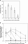
Quantitative analysis of murine lung histopathology during M. pneumoniae infection. A, Mean histopathology score (HPS) for mice (n = 5–8) inoculated with live M. pneumoniae. Time points are at 1, 4, 7, 14, and 35 days postinfection (PI). Values are expressed as mean ± SD. B, Temporal changes in the different inflammatory cell infiltrates are depicted for the above group: a rapid polymorphonuclear and lymphocytic recruitment into the different lung compartments at day 1, pneumonia most severe at day 4 with abundant lymphocytes in vascular and airway walls, loss of pneumonia by day 7, but a persistent presence of lymphoid infiltrates throughout the 35-day study period (see text for details). Open symbols, mice inoculated with SP4 medium; filled symbols, mice inoculated with live M. pneumoniae; open and filled circles, peribronchial/bronchiolar lymphocytes; open and filled diamonds, perivascular lymphocytes; open and filled squares, perivascular polymorphonuclear leukocytes; open and filled triangles, polymorphonuclear leukocytes in alveolar spaces/pneumonia. Values are expressed as mean ± SD.
Figure 3.
Figure 3.
Analysis of murine lung histopathology during M. pneumoniae infection. A, At day 1, there is an abrupt loss of bronchial epithelium at the respiratory bronchiolar level (arrows). The bronchiolar wall contains a pulmonary artery (a) and an inflammatory cellular infiltrate of predominantly lymphocytes with scattered neutrophils (see insert). Conversely, within the alveolar spaces, exudates of numerous neutrophils and fewer alveolar macrophages are evident (circle). (b) indicates bronchiole. Original magnification 20×; insert 40×. B, At day 4, abundant layers of small lymphocytes are aggregated around large and small airways and pulmonary vessels. In the subjacent alveoli, intra-alveolar exudates of neutrophils, nuclear fragments, and alveolar macrophages are evident (circle). Original magnification 20×. C, At day 7, perivascular and peribronchiolar collections of lymphocytes are still present, but as shown in this perivascular site, focal collections of lymphocytes have more cytoplasm and larger nuclei with a more lymphoblastic appearance (left side of insert rectangles; small lymphocytes on right). Alveolar exudate is absent in this site. Original magnifications 20× and 60×. D, At day 35, a few residual foci of lymphocytic infiltrates persist, usually only in perivascular sites and more rarely in both perivascular and peribronchiolar sites as depicted here. Control lungs at this same time period lack appreciable collections of lymphocytes. Original magnification 10×. Tissue sections were stained with hematoxylin and eosin.
Figure 4.
Figure 4.
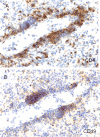
Immunohistochemical characterization of infiltrating lymphocytes associated with perivascular inflamed region of M. pneumoniae–infected mouse lung at day 4 postinfection. The perivascular inflamed site reveals the presence of both CD4+ cells (A) and CD19+ cells (B). Note the distinct and separate aggregation of the 2 lymphocyte populations in each micrograph. Original magnification 20×.
Figure 5.
Figure 5.
Localization of M. pneumoniae and CARDS toxin in the ciliated bronchiolar epithelial region of mouse lung at day 4 postinfection. To localize M. pneumoniae, lung samples were prepared as described in Methods and treated with rabbit anti–M. pneumoniae (1:1500 dilution) and affinity-purified rabbit anti–rCARDS toxin (1:1000 dilution) antisera followed by Vector ABC and diaminobenzidine chromogen. A, Sterile SP4 broth–inoculated control mouse lung with anti–M. pneumoniae antibodies. B, M. pneumoniae–infected mouse lung with anti–M. pneumoniae antibodies. C, Sterile SP4–inoculated mouse lung with anti–rCARDS toxin antibodies. D, M. pneumoniae–infected mouse lung with anti–rCARDS toxin antibodies. Arrows indicate immunostained aggregates of mycoplasma organisms (B) and CARDS toxin (D) that are intermeshed with remaining cilia along the surface of epithelium. Original magnification 60×.
Figure 6.
Figure 6.
Localization of M. pneumoniae cells and CARDS toxin in the alveolar spaces of infected lungs at day 4 postinfection. Cellular exudates contain alveolar macrophages and neutrophils, both of which show immunostained intracytoplasmic aggregates. A, Although more background staining is evident in the cytoplasm of lung cells treated with anti–M. pneumoniae rabbit antiserum, the positively labeled cytoplasmic aggregates are similar in size and distribution with those more easily visualized in the cytoplasm of comparable cell types in the CARDS toxin antibody-stained preparation (B). Samples were processed as described in Figure 5. Original magnification 60×.
Figure 7.
Figure 7.
Seroconversion against CARDS toxin and P1 adhesin in infected mice. Sera were collected from M. pneumoniae–infected and sterile SP4 broth–inoculated mice and analyzed against equimolar concentrations of rCARDS toxin and rP1 adhesin at 1:200 serum dilutions. Immunoglobulin (Ig) M and IgG responses were determined using specific secondary antibodies as described in Methods. Filled squares and circles, serum responses of infected mice to CARDS toxin and P1, respectively; open squares and circles, sterile SP4–inoculated sample background sera responses.

References

    1. Baseman JB, Reddy SP, Dallo SF. Interplay between mycoplasma surface proteins, airway cells, and the protean manifestations of mycoplasma-mediated human infections. Am J Respir Crit Care Med. 1996;154:S137–44. - PubMed
    1. Baseman JB, Tully JG. Mycoplasmas: sophisticated, reemerging, and burdened by their notoriety. Emerg Infect Dis. 1997;3:21–32. - PMC - PubMed
    1. Waites KB, Talkington DF. Mycoplasma pneumoniae and its role as a human pathogen. Clin Microbiol Rev. 2004;17:697–728. - PMC - PubMed
    1. Kraft M, Cassell GH, Henson JE, et al. Detection of Mycoplasma pneumoniae in the airways of adults with chronic asthma. Am J Respir Crit Care Med. 1998;158:998–1001. - PubMed
    1. Nisar N, Guleria R, Kumar S, Chand Chawla T, Ranjan Biswas N. Mycoplasma pneumoniae and its role in asthma. Postgrad Med J. 2007;83:100–4. - PMC - PubMed

Publication types

MeSH terms