Review
. 2012 Jan 10;86(1):1-7.
doi: 10.1095/biolreprod.111.094367. Print 2012 Jan.

Meiotic origins of maternal age-related aneuploidy

Affiliations
Review

Meiotic origins of maternal age-related aneuploidy

Teresa Chiang et al. Biol Reprod. .

Abstract

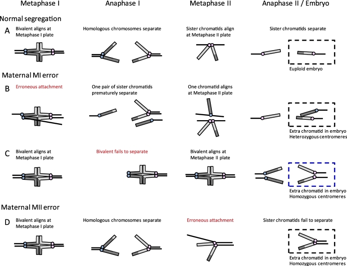
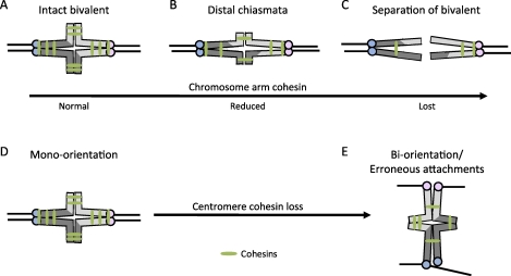
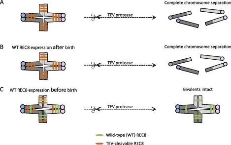
Chromosome segregation errors in female meiosis lead to aneuploidy in the resulting egg and embryo, making them one of the leading genetic causes of spontaneous abortions and developmental disabilities in humans. It is known that aneuploidy of meiotic origin increases dramatically as women age, and current evidence suggests that most errors occur in meiosis I. Several hypotheses regarding the cause of maternal age-related aneuploidy have been proposed, including recombination errors in early meiosis, a defective spindle assembly checkpoint in meiosis I, and deterioration of sister chromatid cohesion with age. This review discusses findings in each area, and focuses especially on recent studies suggesting that deterioration of cohesion with increasing maternal age is a leading cause of age-related aneuploidy.


Figures

FIG. 1.
FIG. 1.
Analysis of centromeres to determine the origin of maternal errors in trisomies. Schematic of chromosome segregation in MI and MII. Chromosomes are depicted as telocentric (as in mice) for simplicity, and centromeres of sister chromatids are colored alike (e.g., blue or pink). A) Normal segregation at both MI and MII, with a euploid embryo. B) Example of an error at MI, where a pair of sister kinetochores erroneously biorients at metaphase I and prematurely separates at anaphase I; the resulting chromatids in the embryo have heterozygous centromeres. Note that there are several possible segregation errors that can occur at MI (e.g., because of failure of recombination or loss of cohesion), of which one example is shown, but heterozygous centromeres in the embryo indicate that an MI error occurred. C) Example of an error at MI, where a bivalent fails to separate at anaphase I and instead separates at anaphase II. Here the centromeres in the embryo are homozygous and would be problematically classified as a MII error, even though the initial error occurred in MI. D) Example of an MII error, where sister chromatids fail to separate at anaphase II; the resulting chromatids in the embryo are homozygous.
FIG. 2.
FIG. 2.
The effects of reduced chromosome cohesion in MI. AC) Chromosome arm cohesion distal to crossover sites holds bivalents together in MI (A). Loss of arm cohesion leads to a shift of chiasmata toward the distal end of chromosomes (B), and complete loss of arm cohesion results in premature complete separation of a bivalent (C). DE) Centromere cohesion holds sister kinetochores together to facilitate mono-orientation (D), and a reduction of centromere cohesion promotes sister kinetochores to erroneously biorient at MI (E).
FIG. 3.
FIG. 3.
Schematic of experiments to test when functional chromosome cohesion is established. Transgenic mice with cleavage sites specific for TEV protease on REC8 were engineered [64]. A) Microinjecting TEV protease into oocytes where bivalents were held together by TEV-cleavable REC8 resulted in complete bivalent and chromatid separation. B) When a transgene encoding Myc-tagged wild-type REC8 (WT REC8) was expressed after birth, the introduction of TEV protease also led to complete chromosome separation. C) When WT REC8 was expressed in oocytes before birth, bivalents remained intact even after microinjection of TEV protease.

References

    1. Penrose LS. The relative effects of paternal and maternal age in mongolism. J Genet 1933; 88: 9 14 - PubMed
    1. Lejeune J, Gautier M, Turpin R. [Study of somatic chromosomes from 9 mongoloid children]. C R Hebd Seances Acad Sci 1959; 248: 1721 1722 - PubMed
    1. Hassold T, Hunt P. To err (meiotically) is human: the genesis of human aneuploidy. Nat Rev Genet 2001; 2: 280 291 - PubMed
    1. Hassold T, Hall H, Hunt P. The origin of human aneuploidy: where we have been, where we are going. Hum Mol Genet 2007; 16 (spec no. 2): R203 R208 - PubMed
    1. May KM, Jacobs PA, Lee M, Ratcliffe S, Robinson A, Nielsen J, Hassold TJ. The parental origin of the extra X chromosome in 47, XXX females. Am J Hum Genet 1990; 46: 754 761 - PMC - PubMed

Publication types