. 2011 Dec;85(23):12304-14.
doi: 10.1128/JVI.05691-11. Epub 2011 Sep 28.

Molecular determinants of arenavirus Z protein homo-oligomerization and L polymerase binding

Affiliations

Molecular determinants of arenavirus Z protein homo-oligomerization and L polymerase binding

Maria Eugenia Loureiro et al. J Virol. 2011 Dec.

Abstract

The arenavirus Z is a zinc-binding RING protein that has been implicated in multiple functions during the viral life cycle. These roles of Z involve interactions with viral and cellular proteins that remain incompletely understood. In this regard, Z inhibits viral RNA transcription and replication through direct interaction with the viral L polymerase. Here, we defined the L-binding domain of Tacaribe virus (TCRV) Z protein and the structural requirements mediating Z homo-oligomerization. By using site-directed mutagenesis, coimmunoprecipitation, and functional assays, we showed that residues R37, N39, W44, L50, and Y57, located around the zinc coordination site I, play a critical role in the Z-L interaction. We also found that Z protein from either TCRV or the pathogenic Junin virus (JUNV) self-associates into oligomeric forms in mammalian cells. Importantly, mutation of the myristoylation site, the strictly conserved residue G at position 2, severely impaired the ability of both TCRV Z and JUNV Z to self-interact as well as their capacity to accumulate at the plasma membrane, strongly suggesting that Z homo-oligomerization is associated with its myristoylation and cell membrane targeting. In contrast, disruption of the RING structure or substitution of W44 or N39, which are critical for L protein recognition, did not affect Z self-binding. Overall, the data presented here indicate that homo-oligomerization is not a requirement for Z-L interaction or Z-mediated polymerase activity inhibition.


Figures

Fig. 1.
Fig. 1.
Effect of deletions in TCRV Z on L binding and Z-mediated reporter gene expression inhibition. CV1 cell monolayers were transfected to express TCRV L protein (+), along with gstZ (lane 1) or each of the Z mutant proteins fused to GST, as indicated on top (lanes 2 to 4 and 6 to 8). As a control, L was coexpressed with GST (lanes 5 and 9). Cells were radiolabeled and lysed as indicated in Materials and Methods. Aliquots of the cell extracts were immunoprecipitated using serum against GST (α-GST panels) or L (α-L panels). The immunoprecipitated proteins were resolved by SDS-PAGE. Z-L binding values (Z-L%), calculated as indicated in Materials and Methods, correspond to the mean of three independent experiments, with relative intrasample variability ranging from 1% (lane 2) to 12% (lane 3). For determination of CAT activity (CAT%), vTF7-3-infected CV1 cell monolayers were transfected with plasmids expressing a TCRV minigenome RNA and TCRV N and L proteins (pWT, pN, and pL, respectively), along with plasmids expressing either GST, gstZ, or each of the indicated Z mutants, as previously described (23). CAT activities in transfected-cell lysates were quantified; mean values of three independent experiments are shown, with relative intrasample variability ranging from 2% (lane 2) to 14% (lane 4). Molecular masses of markers are indicated on the right of each panel. α, anti.
Fig. 2.
Fig. 2.
Mapping of L-binding sites on the TCRV Z region comprising residues 36 to 81. (A) Multiple-sequence alignment of arenavirus Z protein sequences (Clustal method). The region shown corresponds to residues 36 to 81 of TCRV Z protein. GenBank accession numbers of the Z protein sequences used are as follows: NP_694847.1 (TCRV), AAT40445.1 (JUNV), NP_899214.1 (Machupo virus [MACV]), YP_089659.1 (Sabia virus [SABV]), YP_138535.1 (Pichinde virus [PICV]), AAX49343.1 (LCMV), and AAO59510.1 (LASV). Strictly conserved amino acids are marked with asterisks. The positions within the TCRV Z sequence that were mutated are indicated on top with arrowheads. Amino acids of the RING motif are shadowed. (B and C) CV1 cell monolayers were transfected with pL along with either pgstZ(36-85) (B, lanes 1, 10, 16, and 19), pgstZ (C, lanes 1, 9, and 14), the plasmids expressing each of the gstZ(36-85) mutants (B, lanes 2 to 5, 7 to 9, 12 to 15, and 18), or those expressing gstZ mutants (C, lanes 2 to 4, 6 to 8, and 11 to 13), as indicated. In lanes 6, 11, 17, and 20 (B) and lanes 5 and 10 (C), pL was cotransfected with the GST-expressing plasmid. Aliquots of the cell lysates were immunoprecipitated using monospecific serum against either GST (α-GST, top panels) or L (α-L, bottom panels). The immunoprecipitated proteins were resolved by SDS-PAGE, and Z-L binding (Z-L%) was calculated as described in Materials and Methods. Mean values of three independent experiments are shown; standard deviations ranged from 4 to 10%.
Fig. 3.
Fig. 3.
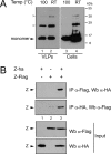
Z protein self-interacts. (A) BSR cells expressing TCRV Z-ha were lysed at 48 h posttransfection in nonreducing SDS-PAGE sample buffer (Invitrogen), and VLPs were purified from the culture supernatants, as described before (8). After addition of β-mercaptoethanol, aliquots corresponding to about 1 × 10⁴ to 5 × 10⁴ cells from both cell extracts and VLPs were incubated at either 100°C or room temperature (RT) for 5 min and then subjected to SDS-PAGE. Resolved proteins were analyzed by Western blotting using anti-HA antibody. The molecular masses of the markers are indicated on the right. (B) CV1 cells were transfected with plasmids pTCRV Z-ha and pTCRV Z-Flag, either separately (lanes 1 and 2) or together (lane 3), as indicated. Cell extracts, obtained at 24 h posttransfection, were analyzed by Western blotting (Wb) with the indicated antibody either before (Input) or after being immunoprecipitated with anti-Flag (IP α-Flag) or anti-HA (IP α-HA) antibody.
Fig. 4.
Fig. 4.
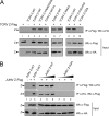
Effect of substitutions in Z on Z-self interaction. (A) CV1 cells were transfected with pTCRV Z-Flag (+) along with the plasmids expressing TCRV Z-ha (TCRV Z-WT) (lanes 3, 6, and 8) or each of the indicated TCRV Z-ha mutants (lanes 4, 5, 7, 9, and 10). As a control, pTCRV Z-Flag (lane 1) and pTCRV Z-ha (lane 2) were transfected alone. (B) CV1 cell monolayers were transfected with pJUNV Z-Flag (+) along with 120 ng (lanes 4 and 7), 250 ng (lanes 5 and 8), or 500 ng (lanes 6 and 9) of the plasmids encoding either wild-type JUNV Z-ha (JUNV Z-WT) or mutant JUNV Z-G2A, as shown. Control monolayers were transfected with pJUNV Z-Flag (lane 2) or the JUNV Z-WT-expressing plasmid (lane 1) alone. (A and B) Aliquots of the cell extracts were analyzed by Western blotting (Wb) with the indicated antibody, either before (Input) or after being immunoprecipitated with anti-Flag (IP α-Flag) or anti-HA (IP α-HA) antibody, as indicated.
Fig. 5.
Fig. 5.
Mutation of residue G2 to alanine affects self-association and intracellular localization of Z protein. (A and B) Analysis of Z-Z interaction by chemical cross-linking. CV1 cells were transfected with plasmids encoding TCRV Z-ha (TCRV Z-WT; A, lanes 1 to 4; B, lanes 1 and 5), TCRV Z-G2A (A, lanes 5 to 8), JUNV Z-ha (JUNV Z-WT; A, lane 9), JUNV Z-G2A (A, lane 10), or each of the mutant TCRV Z-ha proteins indicated in panel B. Cells were incubated with DSP at the concentrations indicated below each panel, as described in Materials and Methods. Aliquots of the DSP-treated cell lysates were incubated for 5 min at 100°C with (B, lane 5) or without (A, lanes 1 to 10; B, lanes 1 to 4) β-mercaptoethanol (βM-OH) and then resolved on SDS-polyacrylamide gels and analyzed by Western blotting, using anti-HA antibody. Bands consistent with monomeric (I), dimeric (II), trimeric (III), or tetrameric (IV) forms of Z are indicated. The molecular masses of markers are indicated on the right of each panel. (C) Intracellular distribution of Z-WT and mutant Z-G2A proteins. BSR cells expressing TCRV Z-WT (frame A), TCRV Z-G2A (frame B), JUNV Z-WT (frame C), or JUNV Z-G2A (frame D) were fixed, and the HA-tagged Z proteins were stained by immunofluorescence and visualized by confocal microscopy as indicated in Materials and Methods. (D) Mutant Z-G2A mediates wild-type levels of polymerase activity inhibition. BSR cells grown in 12-well plates were transfected with 1 μg of pCMV-T7pol, 3 μg of pWT, 3 μg of pTacV N, and 1 μg of pTacV L. When indicated, 0.15 μg of either pTCRV Z-ha (Z-WT) or the plasmids encoding each of the mutant TCRV Z proteins was added to the transfection mix. Cell lysates, obtained at 48 h posttransfection, were used to assay CAT activity (CAT%), as indicated in Materials and Methods. Values correspond to the mean of two independent experiments; relative intrasample variability ranged from 1% to 4%. MAc, monoacetylated chloramphenicol; NAc, nonacetylated chloramphenicol.
Fig. 6.
Fig. 6.
Modeling the L-binding domain of TCRV Z. Ribbon representation of the 3D model of the region spanning residues 36 to 81 of TCRV Z protein (orange), superimposed on the LASV Z structure (gray; PDB accession no. 2KO5). Zinc atoms are shown as light blue spheres. Relevant residues are displayed in stick mode. In the 3D model, residues predicted to be exposed are shown in yellow.

References

    1. Abramoff M. D., Magelhaes P. J., Ram S. J. 2004. Image processing with Image J. Biophotonics Int. 11:36–42
    1. Borden K. L., Campbell Dwyer E. J., Salvato M. S. 1998. An arenavirus RING (zinc-binding) protein binds the oncoprotein promyelocyte leukemia protein (PML) and relocates PML nuclear bodies to the cytoplasm. J. Virol. 72:758–766 - PMC - PubMed
    1. Borden K. L., Campbell Dwyer E. J., Carlile G. W., Djavani M., Salvato M. S. 1998. Two RING finger proteins, the oncoprotein PML and the arenavirus Z protein, colocalize with the nuclear fraction of the ribosomal P proteins. J. Virol. 72:3819–3826 - PMC - PubMed
    1. Buchmeier M. J., de La Torre J. C., Peters C. J. 2007. Arenaviridae: the viruses and their replication, p. 1791–1828 In Knipe D. M., et al. (ed.), Fields virology, 5th ed Lippincott Williams & Wilkins, Philadelphia, PA
    1. Campbell Dwyer E. J., Lai H., MacDonald R. C., Salvato M. S., Borden K. L. 2000. The lymphocytic choriomeningitis virus RING protein Z associates with eukaryotic initiation factor 4E and selectively represses translation in a RING-dependent manner. J. Virol. 74:3293–3300 - PMC - PubMed

Publication types

MeSH terms

LinkOut - more resources