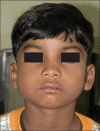
Non-familial cherubism
- PMID: 21957392
- PMCID: PMC3180837
- DOI: 10.4103/0976-237X.83076
Non-familial cherubism
Abstract
Cherubism is a rare, self-limiting, non-neoplastic fibro-osseous disorder of the jaws, usually seen in pediatric population. It is characterized by painless bilateral swelling of the jaws that gives the patient a typical cherubic appearance. Here, we describe the clinical, radiographic, histologic and computed tomographic features of cherubism in a 6-year-old boy.
Keywords: Cherubism; computed tomography; fibro-osseous disorder; giant cell lesions.
Conflict of interest statement
Figures


References
-
- Meng XM, Yu SF, Yu GY. Clinicopathologic study of 24 cases of cherubism. Int J Oral Maxillofac Surg. 2005;34:350–6. - PubMed
-
- Kanbadakone AR, Kadavigere RV, Hosahalli RR, Bhat SS. Case report: CT features of cherubism. Indian J Radiol Imaging. 2008;18:56–9.
-
- Quan F, Grompe M, Jakobs P, Popovich BW. Spontaneous deletion in the FMR 1 gene in a patient with fragile X syndrome and cherubism. Hum Mol Genet. 1995;4:1681–4. - PubMed
-
- Rattan V, Utreja A, Singh BD, Singh SP. Non familial cherubism- A case report. J Indian Soc Pedod Prev Dent. 1997;15:118–20. - PubMed

