. 2011 Sep 29:8:125.
doi: 10.1186/1742-2094-8-125.

Curcumin is a potent modulator of microglial gene expression and migration

Affiliations

Curcumin is a potent modulator of microglial gene expression and migration

Marcus Karlstetter et al. J Neuroinflammation. .

Abstract

Background: Microglial cells are important effectors of the neuronal innate immune system with a major role in chronic neurodegenerative diseases. Curcumin, a major component of tumeric, alleviates pro-inflammatory activities of these cells by inhibiting nuclear factor kappa B (NFkB) signaling. To study the immuno-modulatory effects of curcumin on a transcriptomic level, DNA-microarray analyses were performed with resting and LPS-challenged microglial cells after short-term treatment with curcumin.

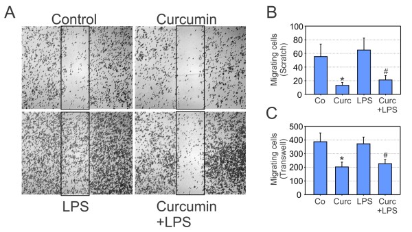
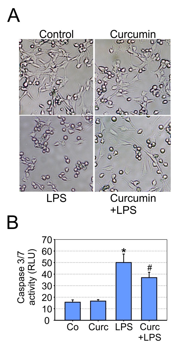
Methods: Resting and LPS-activated BV-2 cells were stimulated with curcumin and genome-wide mRNA expression patterns were determined using DNA-microarrays. Selected qRT-PCR analyses were performed to confirm newly identified curcumin-regulated genes. The migration potential of microglial cells was determined with wound healing assays and transwell migration assays. Microglial neurotoxicity was estimated by morphological analyses and quantification of caspase 3/7 levels in 661W photoreceptors cultured in the presence of microglia-conditioned medium.

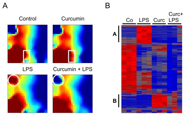
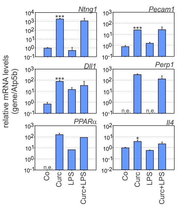
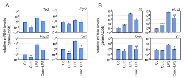
Results: Curcumin treatment markedly changed the microglial transcriptome with 49 differentially expressed transcripts in a combined analysis of resting and activated microglial cells. Curcumin effectively triggered anti-inflammatory signals as shown by induced expression of Interleukin 4 and Peroxisome proliferator activated receptor α. Several novel curcumin-induced genes including Netrin G1, Delta-like 1, Platelet endothelial cell adhesion molecule 1, and Plasma cell endoplasmic reticulum protein 1, have been previously associated with adhesion and cell migration. Consequently, curcumin treatment significantly inhibited basal and activation-induced migration of BV-2 microglia. Curcumin also potently blocked gene expression related to pro-inflammatory activation of resting cells including Toll-like receptor 2 and Prostaglandin-endoperoxide synthase 2. Moreover, transcription of NO synthase 2 and Signal transducer and activator of transcription 1 was reduced in LPS-triggered microglia. These transcriptional changes in curcumin-treated LPS-primed microglia also lead to decreased neurotoxicity with reduced apoptosis of 661W photoreceptor cultures.

Conclusions: Collectively, our results suggest that curcumin is a potent modulator of the microglial transcriptome. Curcumin attenuates microglial migration and triggers a phenotype with anti-inflammatory and neuroprotective properties. Thus, curcumin could be a nutraceutical compound to develop immuno-modulatory and neuroprotective therapies for the treatment of various neurodegenerative disorders.


Figures

Figure 1
Figure 1
Curcumin influences global gene expression in resting and LPS-activated BV-2 microglial cells. (A) Gene Expression Dynamics Inspector (GEDI) analysis of the complete DNA-microarray dataset from control BV-2 cells or cells treated with 20 μM curcumin, 100 ng/ml LPS, or 20 μM curcumin + 100 ng/ml LPS for 6 hours. The white rectangles and circles denote the most prominent expression changes in corresponding gene clusters. (B) Hierarchical clustering of normalized expression levels after one-way ANOVA at p < 0.01. Triplicate microarrays were analyzed for each condition and the pseudo-color scale indicates high (red) or low (blue) expression levels. LPS and curcumin-related clusters are marked with A and B, respectively.
Figure 2
Figure 2
Curcumin induces genes related to adhesion and anti-inflammatory response. Real-time qRT-PCR validation of transcripts in BV-2 microglia stimulated with 20 μM curcumin, 100 ng/ml LPS, or 20 μM curcumin + 100 ng/ml LPS for 6 hours. Relative mRNA levels were quantified for Netrin G1 (Ntng1), Platelet endothelial call adhesion molecule 1 (Pecam1), Delta-like 1 (Dll1), Plasma cell induced endoplasmic reticulum protein 1 (Perp1), Peroxisome proliferator activated receptor α (PPARα), and Interleukin 4 (Il4). Expression was normalized to the control gene Atp5b and mRNA levels (+/- SD) are graphed relative to control cells. Results are calculated from three independent experiments performed in triplicate measurements. * p ≤ 0.05, *** p ≤ 0.001 for curcumin vs. control, Mann-Whitney Rank Sum test. n.e., not expressed.
Figure 3
Figure 3
Curcumin potently blocks pro-inflammatory gene expression. Real-time qRT-PCR validation of transcripts in BV-2 microglia stimulated with 20 μM curcumin, 100 ng/ml LPS, or 20 μM curcumin + 100 ng/ml LPS for 6 hours. Relative mRNA levels were quantified for (A) Toll-like receptor 2 (Tlr2), Early growth response 2 (Egr2), Prostaglandin endoperoxide synthase 2 (Ptgs2), Chemokine ligand 2 (Ccl2), and (B) Interleukin 6 (Il6), Nitric oxide synthase 2 (Nos2, alias iNos), Signal transducer and activator of transcription 1 (Stat1), Complement C3 (C3). Expression was normalized to the control gene Atp5b and mRNA levels (+/- SD) are graphed relative to control cells. Results are calculated from three independent experiments performed in triplicate measurements. * p ≤ 0.05, ** p ≤ 0.01, *** p ≤ 0.001 for curcumin vs. control, and # p ≤ 0.05, ## p ≤ 0.01 for curcumin + LPS vs. LPS, Mann-Whitney Rank Sum test.
Figure 4
Figure 4
Curcumin reduces microglial migration. (A, B) Scratch assays in BV-2 microglia treated with solvent control, 20 μM curcumin, 100 ng/ml LPS, or 20 μM curcumin + 100 ng/ml LPS for 12 hours. (A) Micrographs from one representative experiment out of five independent experiments are shown. (B) Results of scratch assayss are calculated mean values ± SEM from five independent experiments. (C) Transwell chamber migration of BV-2 microglial cells treated with solvent control, 20 μM curcumin, 100 ng/ml LPS, or 20 μM curcumin + 100 ng/ml LPS for 24 hours. The absolute number of migrating cells was counted in the lower chamber and mean values ± SEM are displayed. p ≤ 0.05 for curcumin vs. control, and # p ≤ 0.05 for curcumin + LPS vs. LPS, Student's t test.
Figure 5
Figure 5
Curcumin reduces microglial neurotoxicity on photoreceptors. (A) Phase contrast micrographs showing morphological changes of 661W photoreceptor cell cultures treated with conditioned media from BV-2 cells for 48 hours. The supernatant from control-stimulated, 20 μM curcumin-treated, 100 ng/ml LPS-treated, or 20 μM curcumin + 100 ng/ml LPS-treated cells was added to 661W photoreceptor cells, respectively. The micrographs shown are from one representative experiment out of three independent experiments with the same tendencies. (B) Apoptosis-related caspase 3/7 activation in 661W photoreceptor cells incubated with conditioned media from control-stimulated, 20 μM curcumin-treated, 100 ng/ml LPS-treated, or 20 μM curcumin + 100 ng/ml LPS-treated BV-2 cells. Results are calculated from three independent experiments performed in duplicate measurements. * p ≤ 0.05 for LPS vs. control and # p ≤ 0.05 for curcumin + LPS vs. LPS, respectively, Mann-Whitney Rank Sum test. RLU, relative luciferase units.

References

    1. Hanisch UK, Kettenmann H. Microglia: active sensor and versatile effector cells in the normal and pathologic brain. Nat Neurosci. 2007;10:1387–1394. doi: 10.1038/nn1997. - DOI - PubMed
    1. Streit WJ. Microglia as neuroprotective, immunocompetent cells of the CNS. Glia. 2002;40:133–139. doi: 10.1002/glia.10154. - DOI - PubMed
    1. Giulian D, Li J, Bartel S, Broker J, Li X, Kirkpatrick JB. Cell surface morphology identifies microglia as a distinct class of mononuclear phagocyte. J Neurosci. 1995;15:7712–7726. - PMC - PubMed
    1. Davalos D, Grutzendler J, Yang G, Kim JV, Zuo Y, Jung S, Littman DR, Dustin ML, Gan WB. ATP mediates rapid microglial response to local brain injury in vivo. Nat Neurosci. 2005;8:752–758. doi: 10.1038/nn1472. - DOI - PubMed
    1. Nimmerjahn A, Kirchhoff F, Helmchen F. Resting microglial cells are highly dynamic surveillants of brain parenchyma in vivo. Science. 2005;308:1314–1318. doi: 10.1126/science.1110647. - DOI - PubMed

Publication types