Review
. 2012 Feb;1818(2):272-85.
doi: 10.1016/j.bbamem.2011.09.007. Epub 2011 Sep 16.

Structural correlates of selectivity and inactivation in potassium channels

Affiliations
Review

Structural correlates of selectivity and inactivation in potassium channels

Jason G McCoy et al. Biochim Biophys Acta. 2012 Feb.

Abstract

Potassium channels are involved in a tremendously diverse range of physiological applications requiring distinctly different functional properties. Not surprisingly, the amino acid sequences for these proteins are diverse as well, except for the region that has been ordained the "selectivity filter". The goal of this review is to examine our current understanding of the role of the selectivity filter and regions adjacent to it in specifying selectivity as well as its role in gating/inactivation and possible mechanisms by which these processes are coupled. Our working hypothesis is that an amino acid network behind the filter modulates selectivity in channels with the same signature sequence while at the same time affecting channel inactivation properties. This article is part of a Special Issue entitled: Membrane protein structure and function.


Figures

Figure 1
Figure 1
Topology diagrams of K+ channels colored according to secondary structure location. Transmembrane pore forming helices are cyan, additional transmembrane helices are tan or brown, the helix within the P-loop is pink, and cytosolic helices are green. A. Kv subunits contain six transmembrane helices. Helices S5 and S6 form the pore while helices S3 and S4 function as a voltage sensor. B. Kir subunits contain two transmembrane pore forming helices as well as an additional N-terminal helix and a variable cytosolic domain that allows the channel to be regulated by different substances. C. KCa subunits are topologically more similar to Kv channels. KCa1.1 channels have an additional transmembrane helix (S0) for a total of seven. KCa2 and KCa 3.1 channels have six transmembrane helices like Kv channels, but also contain a cytosolic domain to bind calmodulin. D. Each KcsA subunit is composed of two transmembrane helices connected by a P-loop. These helices extend into the cytosol to form a structural domain that is generally removed to aid crystallization (not shown).
Figure 2
Figure 2
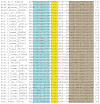
Sequence alignment of representative K+ channels. All sequences except for chicken Kir2.2 were taken from the Uniprot Knowledgebase and are listed with the appropriate accession number. The Kir2.2 sequence was taken from Protein Data Bank entry 3JYC. Sequence regions highlighted in blue or brown represent residues found in the pore helix or helix TM2/S6 based on the structure of KcsA (PDB ID 1K4C). Amino acids highlighted in yellow correspond to the signature sequence residues that make up the selectivity filter. Residues represented as red text are shown/predicted to form interactions within the selectivity filter and/or its surrounding scaffolding as depicted in Figure 3.
Figure 3
Figure 3
Stereo diagram depicting different crytallographically observed KcsA selectivity filter conformations. Two subunits have been removed for clarity. Spheres are colored to represent different species: purple for K+, orange for Na+, and red for H2O. A. Active conformation of filter in presence of high K+ (PDB ID 1K4C). B. Collapsed filter in the presence of Na+ and reduced K+ (PDB ID 1K4D). C. Filter with E71A mutation in the presence of high K+. D80 swings out of the selectivity filter and multiple carbonyls within the filter move out of the conduction pathway. Functionally this mutation removes inactivation. D. Filter with E71A mutation in the absence of K+ and the presence of Na+. The filter looks very similar to the E71A KcsA channel in the presence of high K+ suggesting that the E71A mutation removes inactivation by preventing the selectivity filter from collapsing as in Figure 4B.
Figure 4
Figure 4
Residues involved in KcsA inactivation (F103: orange-space filled, T74 cyan-space filled, D80/E71/W67/V76/M96: yellow-stick, K+: purple sphere, H2O: red sphere). A. Closed structure of KcsA (PDB ID 1K4C) with non-collapsed selectivity filter. B. Open structure of KcsA with collapsed selectivity filter (PDB ID 3F5W). Opening of KcsA via movement of helix TM1 results in contact between F103 (TM1) and T74 (bottom of selectivity filter). In the open structure, the F103 side-chain is shown to rotate to avoid collision with T74. Hydrogen bonds between D80, E71, and W67 have also been linked to inactivation along with interactions between M96 and V76 on a neighboring subunit.
Figure 5
Figure 5
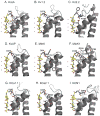
Cartoon diagrams depicting observed/predicted interactions within the selectivity filters and surrounding scaffolding of several K+ channels. Selectivity filter residue carbons are colored yellow and K+ ions are purple spheres. Possible hydrogen bonds are shown as dashed black lines. The first six illustrations were created from the following PDB depositions: A. Kcsa (1K4C). B. Kv1.2 (3LUT). C. Kir2.2 (3JYC). D. KvAP (1ORQ). E. MthK (3LDC). F. MloK1 (3BEH). The final three illustrations: G. KCa1.1. H. KCa2.1. I. HCN1, are homology models as there are no experimentally determined structures available. These homology models were generated in ProtMod using KvAP (PDB 2A0L), Kv1.2 (PDB 2A79), and MloK1 (PDB 2zd9) as the respective starting models [149, 154, 155]. The protein sequence identity and similarity between the initial models and homologous models were 24.6% and 50.9% (KCa1.1), 35.1% and 47.4% (KCa2.1), and 17.5% and 36.8% (HCN1). These values correspond to the 57 residues surrounding the selectivity filter (not the entire protein) and similarity scores were computed using a BLOSUM62 scoring matrix.

Similar articles

Cited by

References

    1. Yu FH, Catterall WA. The VGL-chanome: a protein superfamily specialized for electrical signaling and ionic homeostasis. Sci STKE. 2004;2004:re15. - PubMed
    1. Yellen G. The voltage-gated potassium channels and their relatives. Nature. 2002;419:35–42. - PubMed
    1. Hibino H, Inanobe A, Furutani K, Murakami S, Findlay I, Kurachi Y. Inwardly rectifying potassium channels: their structure, function, and physiological roles. Physiol Rev. 2010;90:291–366. - PubMed
    1. Xia XM, Zeng X, Lingle CJ. Multiple regulatory sites in large-conductance calcium-activated potassium channels. Nature. 2002;418:880–884. - PubMed
    1. Bao L, Rapin AM, Holmstrand EC, Cox DH. Elimination of the BK(Ca) channel’s high-affinity Ca(2+) sensitivity. J Gen Physiol. 2002;120:173–189. - PMC - PubMed

Substances

LinkOut - more resources