Case Reports
. 2011 Sep 29:6:122.
doi: 10.1186/1749-8090-6-122.

A silent gigantic solitary fibrous tumor of the pleura: case report

Affiliations
Case Reports

A silent gigantic solitary fibrous tumor of the pleura: case report

Nobuyuki Furukawa et al. J Cardiothorac Surg. .

Abstract

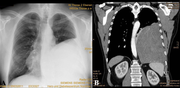
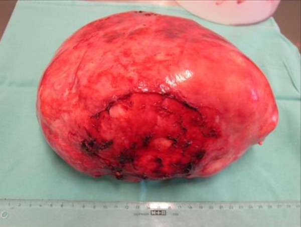
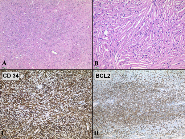
Solitary fibrous tumor of the pleura is a rare mesenchymal tumor, representing less than 5% of all neoplasms associated with the pleura. A 57-year-old man had general malaise without chest symptoms for 1 month. A chest roentgenogram and computed tomography showed a giant mass in the left thorax. Although the tumor compressed the descending aorta and other mediastinal structures strongly, thereby shifting them to the right side, the patient had no symptoms except malaise. The tumor was successfully resected via two separate thoracotomies. The tumor was measured (20 cm × 19 cm × 15 cm) and weighed (2150 g). The tumor was histologically and immunohistochemically diagnosed as benign. Although SFT is benign, a long follow-up period is essential as even patients with complete resection are at risk of recurrence many years after surgery.


Figures

Figure 1
Figure 1
Chest radiography and CT scan images. (A) Initial chest radiography revealed a large well-circumscribed mass in the left thorax. (B) Initial contrast-enhanced computed tomography showed a huge homogeneous, sharply defined mass compressing the aorta.
Figure 2
Figure 2
The gigantic encapsulated solitary fibrous tumor of the pleura, weighed 2150 g and measured 20 cm × 19 cm × 15 cm.
Figure 3
Figure 3
Microscopic examination of solitary fibrous tumor of the pleura. (A, B) Microscopic specimen of the tumor shows solid proliferation of spindle-shaped fibroblastic cells in a patternless pattern. (Hematoxylin and eosin; magnification 40× and 200×) (C, D) Spindle-shaped tumor cells show strong positivities for immunohistochemical staining with CD34 (C) and BCL2 (D).

References

    1. Harrison-Phipps KM, Nichols FC, Schleck CS, Deschamps C, Cassivi SD, Schipper PH, Allen MS, Wigle DA, Pairolero PC. Solitary fibrous tumors of pleura: Results of surgical treatment and long-term prognosis. J Thorac Cardiovasc Surg. 2009;138:19–25. doi: 10.1016/j.jtcvs.2009.01.026. - DOI - PMC - PubMed
    1. Shaker W, Meatchi T, Dusser D, Riquet M. An unusual presentation of solitary fibrous tumor of the pleura: right atrium and inferior vene cava compression. Eur J Cardiothorac Surg. 2002;22:640–2. doi: 10.1016/S1010-7940(02)00432-3. - DOI - PubMed
    1. Cardillo G, Carbone L, Carleo F, Masala N, Graziano P, Bray A, Martelli M. Solitary fibrous tumors of the pleura: An analysis of 110 patients treated in a single institution. Ann Thorac Surg. 2009;88:1632–7. doi: 10.1016/j.athoracsur.2009.07.026. - DOI - PubMed
    1. Hu CK, Chang YL, Lin WC, Lee YC. Resection of giant thoracic solitary fibrous tumor through two separate thoracotomies. J Thorac Cardiovasc Surg. 2008;136:1077–9. doi: 10.1016/j.jtcvs.2007.12.015. - DOI - PubMed
    1. Altinok T, Topcu S, Tastepe I, Yazici Y, Cetin G. Localized fibrous tumors of the pleura: Clinical and surgical evaluation. Ann Thorac Surg. 2003;76:892–5. doi: 10.1016/S0003-4975(03)00663-5. - DOI - PubMed

Publication types

LinkOut - more resources