. 2011 Sep 29;2(1):11.
doi: 10.1186/1759-8753-2-11.

Retrotransposition of R2 elements in somatic nuclei during the early development of Drosophila

Affiliations

Retrotransposition of R2 elements in somatic nuclei during the early development of Drosophila

Michael T Eickbush et al. Mob DNA. .

Abstract

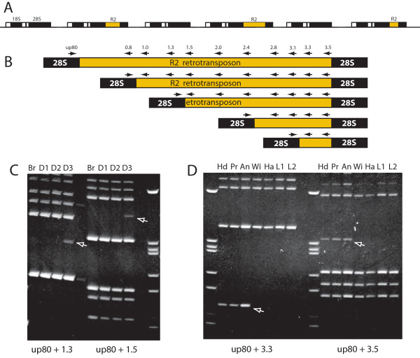
Background: R2 retrotransposable elements exclusively insert in the 28S rRNA genes of their host. Their RNA transcripts are produced by self-processing from a 28S R2 cotranscript. Because full-length R2 transcripts are found in most tissues of R2-active animals, we tested whether new R2 insertions occurred in somatic tissues even though such events would be an evolutionary dead end.

Findings: PCR assays were used to identify somatic R2 insertions in isolated adult tissues and larval imaginal discs of Drosophila simulans. R2 somatic mosaics were detected encompassing cells from individual tissues as well as tissues from multiple body segments. The somatic insertions had 5' junction sequences characteristic of germline insertions suggesting they represented authentic retrotransposition events.

Conclusions: Body segments are specified early in Drosophila development, thus the detection of the same somatic insertion in cells from multiple tissues suggested that the R2 retrotransposition events had occurred before the blastoderm stage of Drosophila development. R2 activity at this stage, when embryonic nuclei are rapidly dividing in a common cytoplasm, suggests that some retrotransposition events appearing as germline events may correspond to germline mosaicism.


Figures

Figure 1
Figure 1
Diagram of R2 insertions within the rRNA genes of Drosophila and the PCR assay used to monitor somatic mosaicism. (A) Diagram of the tandemly repeated rRNA genes of Drosophila simulans and the location of R2 insertions. Black boxes, 18S, 5.8S and 28s rRNA genes (5.8S gene between the 18S and 28S genes is not labeled); white boxes, transcribed spacer regions. (B) About half of the R2 insertions have deletions of their 5' end that can extend to nearly the entire length of the element. All R2 copies have the same 3' junction with the 28S gene. Arrows above the R2/28S diagrams indicate the positions of the oligonucleotide primers used to assay for the 5' truncations. The DNA extraction method, the primers used and the PCR protocols used were identical to those in previous reports [11-13]. (C) Examples of the ethidium stained PCR products derived from larval tissues. The larval tissues were dissected in Drosophila Ringers. (D) Examples of the ethidium stained PCR products derived from adult tissues. Adult tissues were placed directly in the DNA extraction solution. PCR bands interpreted as somatic insertions are indicated with arrows. To be scored as a somatic mosaic the amplified band had to be detected with two sets of primer combinations (shown below the figures). The following abbreviations for body segments were used: An = antenna; Br, brain from a larvae; D1-D3, individual pairs of imaginal disc (the specific disc pair used was not known); Ha = haltere; Hd = head; L1 and L2 = individuals legs from different body segments; Pr = proboscis; Wi = wing.
Figure 2
Figure 2
Diagram of the 5' junction sequences of the somatic R2 insertions with the 28S gene. Putative somatic insertions such as those shown in Figure 1C, D were excised from a gel, re-amplified with the same PCR primers, purified on a second gel and subjected to double-stranded DNA sequencing. The 28S gene sequence is shown at the top of the figure. Short regions upstream and downstream of the R2 insertion site (arrow) are not shown because no R2 junctions occurred in these areas. For each junction those sequences corresponding to R2 sequences have been highlighted with tan shaded boxes. Those nucleotides that could correspond to either the 28S sequence or R2 (described as microidentities in the text) have been indicated with a blue box. Those sequences that do not correspond to either the 28S gene or R2 (described as non-templated nucleotides in the text) are indicated with an orange box. The number at each junction corresponds to the first nucleotide of the R2 element, based on the consensus Drosophila simulans R2 sequence [18].

References

    1. Laski FA, Rio DC, Rubin GM. Tissue specificity of Drosophila P element transposition is regulated at the level of mRNA splicing. Cell. 1986;44:7–19. doi: 10.1016/0092-8674(86)90480-0. - DOI - PubMed
    1. Chaboissier MC, Busseau I, Prosser J, Finnegan DJ, Bucheton A. Identification of a potential RNA intermediate for transposition of the LINE-like element I factor in Drosophila melanogaster. EMBO J. 1990;9:3557–3563. - PMC - PubMed
    1. Emmons SW, Yesner L. High-frequency excision of transposable element Tc1 in the nematode Caenorhabditis elegans is limited to somatic cells. Cell. 1984;36:599–605. doi: 10.1016/0092-8674(84)90339-8. - DOI - PubMed
    1. van den Hurk JA, Meij IC, Seleme MC, Kano H, Nikopoulos K, Hoefsloot LH, Sistermans EA, de Wijs IJ, Mukhopadhyay A, Plomp AS, de Jong PTVM, Kazazian HH Jr, Cremers FPM. L1 retrotransposition can occur early in human embryonic development. Hum Mol Genet. 2007;16:1587–1592. doi: 10.1093/hmg/ddm108. - DOI - PubMed
    1. Kano H, Godoy I, Courtney C, Vetter MR, Gerton GL, Ostertag EM, Kazazian HH Jr. L1 retrotransposition occurs mainly in embryogenesis and creates somatic mosaicism. Genes Dev. 2009;23:1303–1312. doi: 10.1101/gad.1803909. - DOI - PMC - PubMed

LinkOut - more resources