Review
. 2011 Sep 27;21(18):R750-7.
doi: 10.1016/j.cub.2011.06.033.

Coordinating growth and maturation - insights from Drosophila

Affiliations
Review

Coordinating growth and maturation - insights from Drosophila

Jason M Tennessen et al. Curr Biol. .

Abstract

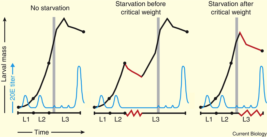
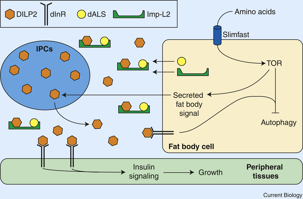
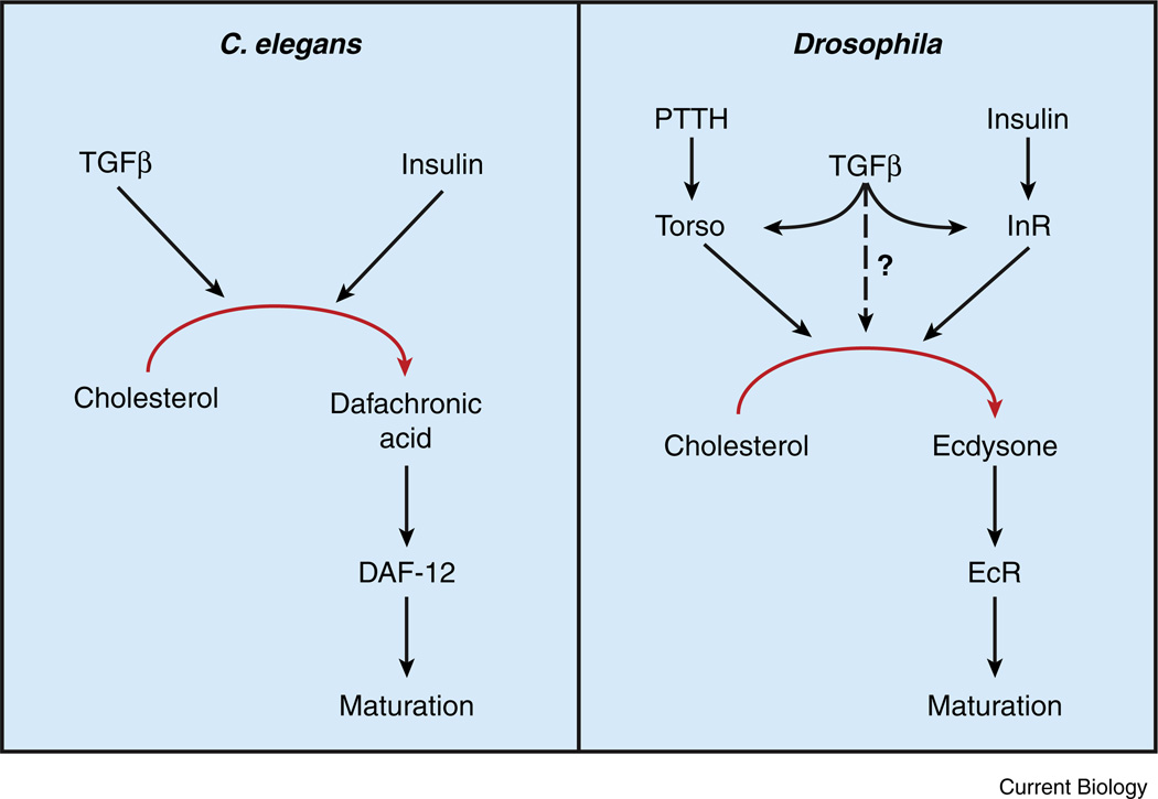
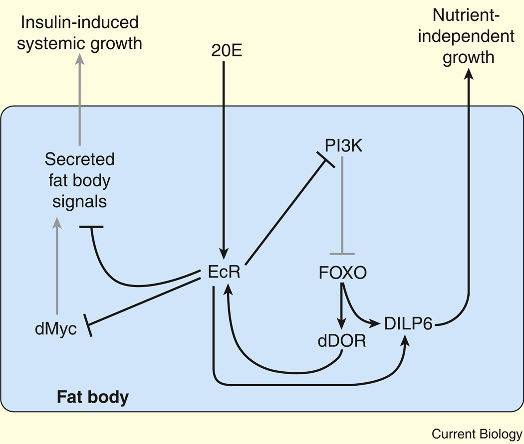
Adult body size in higher animals is dependent on the amount of growth that occurs during the juvenile stage. The duration of juvenile development, therefore, must be flexible and responsive to environmental conditions. When immature animals experience environmental stresses such as malnutrition or disease, maturation can be delayed until conditions improve and normal growth can resume. In contrast, when animals are raised under ideal conditions that promote rapid growth, internal checkpoints ensure that maturation does not occur until juvenile development is complete. Although the mechanisms that regulate growth and gate the onset of maturation have been investigated for decades, the emerging links between childhood obesity, early onset puberty, and adult metabolic disease have placed a new emphasis on this field. Remarkably, genetic studies in the fruit fly Drosophila melanogaster have shown that the central regulatory pathways that control growth and the timing of sexual maturation are conserved through evolution, and suggest that this aspect of animal life history is regulated by a common genetic architecture. This review focuses on these conserved mechanisms and highlights recent studies that explore how Drosophila coordinates developmental growth with environmental conditions.


Figures

Figure 1
Figure 1. A schematic representation of Drosophila larval growth and development
Drosophila larvae experience exponential growth (black line) as they develop through three distinct larval instars (L1, L2, and L3). Pulses of 20E (blue line) direct progression through the larval molts. The critical weight checkpoint (grey vertical line) occurs near the L2–L3 molt. A series of low-titer 20E pulses occur at ~8, 20, and 28 h after the L2–L3 molt, followed by a high-titer 20E pulse at the end of L3 that triggers puparium formation [7]. If an animal is starved (red lines) prior to the attainment of critical weight, development stalls until the larva finds a new food source, but final body size is unaffected. After critical weight is achieved, starvation inhibits growth but no longer affects developmental progression, resulting in a significantly smaller final body size.
Figure 2
Figure 2. The larval fat body regulates systemic growth
Pre-critical weight larval growth is regulated by nutrient-dependent signals that emanate from the fat body. Ingested amino acids are sensed by fat body cells and activate TOR kinase, which promotes the release of an unknown factor that stimulates DILP2 secretion from the insulin-producing cells (IPCs). DILP2, in turn, promotes growth and development in peripheral tissues by binding to the insulin receptor (dInR) and activating the insulin signaling pathway. Additionally, the fat body releasesd ALS and Imp-L2, which form a stable complex with DILP2 and dampen insulin signaling.
Figure 3
Figure 3. A conserved genetic hierarchy regulates animal maturation
A combination of insulin and TGFβ signaling regulates steroid hormone production and maturation in both C. elegans and Drosophila. In worms, dietary nutrients and favorable growth conditions increase TGFβ and insulin signaling in endocrine tissues and stimulate dafachronic acid (DA) synthesis. DA systemically activates the nuclear receptor DAF-12, thereby preventing dauer formation and promoting maturation. In Drosophila, TGFβ signaling in the prothoracic gland (PG) upregulates expression of Torso and the insulin receptor (dInR), which promote ecdysone synthesis in response to PTTH and insulin, respectively. Ecdysone is then released from the PG, converted into 20E, and promotes maturation by systemically activating the ecdysone receptor (EcR).
Figure 4
Figure 4. Ecdysone functions in the fat body to regulate systemic growth
EcR activation by 20E in the fat body inhibits systemic insulin signaling and growth, in part by downregulating dMyc expression. EcR also inhibits PI3K signaling, which allows dFOXO to translocate to the nucleus and activate the expression of target genes. These include dDOR and dilp6, which is also upregulated by 20E–EcR. dDOR further activates EcR signaling, while dilp6 promotes nutrient-independent growth. Grey lines represent genetic interactions that are downregulated by 20E signaling.

References

    1. Robertson CW. The metamorphosis of Drosophila melanogaster, including an accurately timed account of the principal morphological changes. J. Morphol. 1936;59:351–399.
    1. Thummel CS. Molecular mechanisms of developmental timing in C. elegans and Drosophila. Dev. Cell. 2001;1:453–465. - PubMed
    1. Riddiford LM, Cherbas P, Truman JW. Ecdysone receptors and their biological actions. Vitam. Horm. 2001;60:1–73. - PubMed
    1. Gilbert LI, Warren JT. A molecular genetic approach to the biosynthesis of the insect steroid molting hormone. Vitam. Horm. 2005;73:31–57. - PubMed
    1. Huang X, Warren JT, Gilbert LI. New players in the regulation of ecdysone biosynthesis. J. Genet. Genomics. 2008;35:1–10. - PubMed

Publication types

LinkOut - more resources