. 2011 Dec;37(4):319-27.
doi: 10.1016/j.jaut.2011.09.003. Epub 2011 Sep 29.

MCAM-expressing CD4(+) T cells in peripheral blood secrete IL-17A and are significantly elevated in inflammatory autoimmune diseases

Affiliations

MCAM-expressing CD4(+) T cells in peripheral blood secrete IL-17A and are significantly elevated in inflammatory autoimmune diseases

Pradeep K Dagur et al. J Autoimmun. 2011 Dec.

Abstract

Th17 cells are a subset of CD4(+) T cells characterized by production of IL-17 and are known to be key participants in inflammatory reactions and various autoimmune diseases. In this study we found that a subset of human CD4(+) T cells expressing MCAM (CD146) have higher mRNA levels of RORC2, IL-23R, IL-26, IL-22, IL-17A, but not IFN-γ, compared to CD4(+) T cell not expressing CD146. Upon TCR stimulation with CD3/CD28, CD4(+)CD146(+) T cells secrete significantly more IL-17A, IL-6, and IL-8 than do CD4(+)CD146(-) T cells. Low frequencies of CD4(+)CD146(+) T cells are found in the circulation of healthy adults, but the frequency of these cells is significantly increased in the circulation of patients with inflammatory autoimmune diseases including Behcet's, sarcoidosis and Crohn's disease. Patterns of gene expression and cytokine secretion in these cells are similar in healthy and disease groups. In Crohn's disease, the increase in CD4(+)CD146(+) cells in the circulation correlates with disease severity scores. These data indicate that expression of CD146 on CD4(+) T cells identifies a population of committed human Th17 cells. It is likely the expression of CD146, an endothelial adhesion molecule, facilitates adherence and migration of Th17 cells through the endothelium to sites of inflammation.


Figures

Figure 1
Figure 1. CD146 expression on CD4+ T-cells
Single-cell suspensions from healthy donors and patients with various inflammatory diseases were stained using a combination of antibodies as described in the material and methods. A. Lymphocytes were identified based on their forward and side scatter properties. Subsequently, CD3 was used to identify T cells (CD3+) among the previously selected lymphocytes. CD4+ T cells and CD8+ T cells were identified as uniquely expressing CD4 or CD8 antigens. CD146+ expression was then determined on the gated CD4+ T cells. B. Representative bivariate plots of CD146 expression in CD4+ T cells (as described above) in healthy, Behcet’s, sarcoidosis and Crohn’s patients. C. The mean percentages (±SEM) of CD4+ T cells expressing CD146 among healthy (n=73), Behcet’s (n=20), sarcoidosis (n=24) and Crohn’s (n=20) patients. D. Correlation between frequency of CD4+CD146+ T cells in peripheral circulation and disease severity scores in Crohn’s disease (Harvey-Bradshaw Index (HBI) [24]. With this index, active disease is defined as a HBI score of ≥4. HBI scores were correlated to the frequency of CD4+CD146+ T cells. Represented is the linear regression between the 2 parameters.
Figure 2
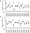
Figure 2. mRNA level of CD4+CD45RO+CD146+ T lymphocytes in circulation compared to CD4+CD45RO+CD146 T cells
A. Single-cell suspensions from healthy donors (n=7) were stained and sorted into 2 populations: CD4+CD45RO+CD146+ (Dark circles) and CD4+CD45RO+CD146 (Open circles). After sorting, mRNA was extracted and RT-PCR was performed using gene expression assays as described in the methods. B. PBMCs were stimulated with PMA/ionomycin for 16hrs as described in the material and methods, then sorted into CD4+CD45RO+CD146+ (Dark circles) and CD4+CD45RO+CD146 (Open circles) subsets. mRNA isolation and RT-PCR, using the same gene expression as described previously, was performed following the sort.
Figure 3
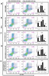
Figure 3. Expression of chemokine receptors and Th17 phenotype-associated markers on CD4+CD146+ T cells
Single-cell suspensions from healthy donors and patients with various inflammatory diseases were stained with a combination of antibodies to study the co-expression of CD146 with other markers associated with a Th17 phenotype. The figure shows representative bivariate dot plots of CD146 co-expression with (A) CD161, (B) CCR-6, (C) CCR-4, (D) CCR-2 and (E) CXCR-3.
Figure 4
Figure 4. Intracellular cytokines in freshly isolated CD4+CD45RO+CD146+ and CD4+CD45RO+CD146 T lymphocytes
PBMCs from healthy individuals (n=7) and patients with various inflammatory diseases (n=4) were stimulated using PMA/ionomycin and stained for intracellular cytokines IL-17A and IFN-γ. Data shown are the percentages of IFN-γ (left panel) and IL-17A (right panel) positive cells in CD4+CD45RO+CD146+ and CD4+CD45RO+CD146 subsets of healthy donors (H) and patients (D).
Figure 5
Figure 5. m-RNA levels in CD4+CD45RO+CD146+ T cells compared to CD4+CD45RO+CD146 T cells after in vitro stimulation
Single-cell suspensions from healthy individuals (n=7) were stained and sorted into 2 populations CD4+CD45RO+CD146+ (Dark circles) and CD4+CD45RO+CD146 (Open circles). These subsets were stimulated with CD3/CD28 as described in the material and methods. mRNA isolation was performed after 5 days of culture. RT-PCR were performed using gene expression assay from Applied Biosystems measuring as described in the methods.
Figure 6
Figure 6. Intracellular cytokine expression of CD4+CD45RO+CD146+ T cells and CD4+CD45RO+CD146 T cells in presence of Th17 polarizing conditions
Single-cell suspensions from healthy individuals (n≥3) were stained and sorted into 2 populations: CD4+CD45RO+CD146+ and CD4+CD45RO+CD146. These sorted populations were subjected to different in vitro stimulations and the expression of IL-17A (x axis) and IFN-γ (y axis) was measured. CD4+CD45RO+CD146 T cells are shown in the left panel and CD4+CD45RO+CD146+ T cells are shown in the center panel, and summary data are shown in the right panel. The condition of stimulations were: (A) plate bound CD3 and CD28 (B) plate bound CD3 and CD28 and IL-1β, (C) plate bound CD3 and CD28 and IL-23, (D) plate bound CD3 and CD28 and IL-1β+IL-23 and (E) plate bound CD3 and CD28 and IL-1β+IL-23+TGF-β+IL-6.
Figure 7
Figure 7. Cytokine secretion of CD4+CD45RO+CD146+ T cells and CD4+CD45RO+CD146 T cells
Single-cell suspensions from healthy individuals (n=7) were stained and sorted into 2 populations CD4+CD45RO+CD146+ and CD4+CD45RO+CD146. These subsets were stimulated with CD3/CD28 as described in the material and methods. Secretion of IL-17A (A), IFN-γ (B), IL-8 (C) and IL-6 (D) were measured in culture supernatant using multiplex bead array assays in CD4+CD146 or CD4+CD146+ subsets.

References

    1. Mosmann TR, Cherwinski H, Bond MW, Giedlin MA, Coffman RL. Two types of murine helper T cell clone. I. Definition according to profiles of lymphokine activities and secreted proteins. Journal of immunology. 1986;136:2348–57. - PubMed
    1. Killar L, MacDonald G, West J, Woods A, Bottomly K. Cloned, Ia-restricted T cells that do not produce interleukin 4(IL 4)/B cell stimulatory factor 1(BSF-1) fail to help antigen-specific B cells. Journal of immunology. 1987;138:1674–9. - PubMed
    1. Sakaguchi S, Sakaguchi N, Asano M, Itoh M, Toda M. Immunologic self-tolerance maintained by activated T cells expressing IL-2 receptor alpha-chains (CD25). Breakdown of a single mechanism of self-tolerance causes various autoimmune diseases. Journal of immunology. 1995;155:1151–64. - PubMed
    1. Bendelac A, Killeen N, Littman DR, Schwartz RH. A subset of CD4+ thymocytes selected by MHC class I molecules. Science. 1994;263:1774–8. - PubMed
    1. Weiner HL. Induction and mechanism of action of transforming growth factor-beta-secreting Th3 regulatory cells. Immunol Rev. 2001;182:207–14. - PubMed

Publication types

MeSH terms