. 2011 Oct 28;12(11):1175-81.
doi: 10.1038/embor.2011.174.

PPM1A dephosphorylates RanBP3 to enable efficient nuclear export of Smad2 and Smad3

Affiliations

PPM1A dephosphorylates RanBP3 to enable efficient nuclear export of Smad2 and Smad3

Fangyan Dai et al. EMBO Rep. .

Abstract

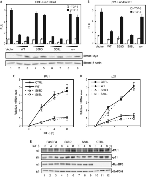
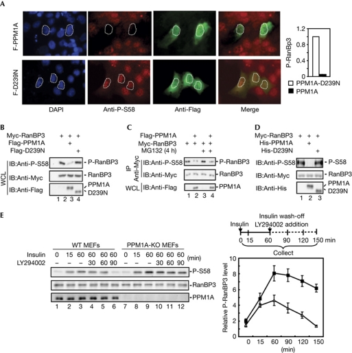
Smad2 and Smad3 (Smad2/3) are essential signal transducers and transcription factors in the canonical transforming growth factor-β (TGF-β) signalling pathway. Active Smad2/3 signalling in the nucleus is terminated by dephosphorylation and subsequent nuclear export of Smad2/3. Here we report that protein phosphatase PPM1A regulates the nuclear export of Smad2/3 through targeting nuclear exporter RanBP3. PPM1A directly interacted with and dephosphorylated RanBP3 at Ser 58 in vitro and in vivo. Consistently, RanBP3 phosphorylation was elevated in PPM1A-null mouse embryonic fibroblasts. Dephosphorylation of RanBP3 at Ser 58 promoted its ability to export Smad2/3 and terminate TGF-β responses. Our findings indicate the critical role of PPM1A in maximizing exporter activity of RanBP3 for efficient termination of canonical TGF-β signalling.


Conflict of interest statement

The authors declare that they have no conflict of interest.

Figures

Figure 1
Figure 1
S58D substitution impairs the inhibitory function of RanBP3 in transforming growth factor-β (TGF-β) signalling. (A) Effect of RanBP3 phosphorylation mutants on SBE-Luc response. HaCaT cells were transfected with indicated plasmids and treated with 2 ng/μl TGF-β for 20 h, and cell lysates were subjected to reporter assays. Values and error bars represent average and standard deviation of three independent experiments. The expression level of RanBP3 (wild type, WT) and its mutants were examined by western blotting using Myc antibody (bottom). (B) Effect of RanBP3 phosphorylation mutants on the natural p21 promoter (p21-Luc) activity in HaCaT cells. Values and error bars represent average and standard deviation of four independent experiments. (C) Quantitative real-time reverse transcription–polymerase chain reaction (qRT–PCR) analysis of p21 messenger RNA (mRNA). HaCaT cells stably expressing Flag-tagged RanBP3 (WT, S58D, S58L) and parental HaCaT cells (CTRL) were treated with TGF-β (2 ng/μl) for up to 8 h, and were subjected to total RNA extraction. Values and error bars represent average and standard deviation of three independent experiments. (D) qRT–PCR analysis of PAI1 mRNA. Values and error bars represent average and standard deviation of three independent experiments. (E) Western blotting analysis of p21 and PAI1. HaCaT cells stably expressing Flag-tagged RanBP3 (WT, S58D, S58L) and parental HaCaT cells (CTRL) were treated with TGF-β (2 ng/μl) for 8 h and cell lysates collected at indicated times. The levels of p21, Plasminogen activator inhibitor 1 (PAI1) and RanBP3 proteins were examined by western blotting using indicated antibodies. The glyceraldehyde-3-phosphate dehydrogenase (GAPDH) blot serves as a loading control. SBE, Smad-binding element; RLU, Relative luciferase unit; wv, RanBP3 Ran-binding mutant.
Figure 2
Figure 2
RanBP3-S58D fails to promote nuclear export of Smad2/3. (A) Microscopic analysis of Smad2/3 nuclear accumulation. HaCaT cells were transfected with Myc-tagged RanBP3 or mutants. After 24 h transfection, cells were treated with transforming growth factor-β (TGF-β, 2 ng/μl) for 2 h and fixed. Endogenous Smad2/3 (red) was immunostained by using anti-Smad2 (left) or Smad3 (right) antibody, respectively. The arrow indicates the cells with ectopically expressed Myc-RanBP3 or its mutants detected by anti-Myc immunostaining (green). 4,6-Diamidino-2-phenylindole (DAPI) stains the nucleus (blue). Intensity of nuclear Smad3 among these cells was quantified using National Institutes of Health Image software. Far right panel shows the percentage of cells with decreased nuclear Smad3 level (red bar) within 24 randomly counted cells. (B) Western blot analysis of Smad2/3 export. Cells were collected at the indicated times (depicted on the right) and the cytoplasmic/nuclear fractions were extracted. Successful fractionation was confirmed by western blot analysis of the cytoplasmic marker glyceraldehyde-3-phosphate dehydrogenase (GAPDH) and nuclear marker Lamin A/C. Smad2/3 in the cytoplasmic or nuclear fractions were examined by anti-Smad2/3 western blotting. SB431542 is a TGF-β type I receptor inhibitor. (C) Quantitative analysis of Smad2 export. Details are described in Methods. Values and error bars represent average and standard deviation of three independent experiments. The expression level of RanBP3 and mutants were examined by western blot (bottom). The β-actin blot serves as a loading control. (D) In vitro import assay of Smad2 import in permeabilized cells. Details are described in Methods. Nuclear imported Smad2 was examined by western blot using anti-Smad2 antibody. The Lamin A/C blot serves as a nuclear marker and loading control. WT, wild type.
Figure 3
Figure 3
PPM1A dephosphorylates RanBP3 at Ser 58. (A) Microscopic analysis of PPM1A phosphatase activity on RanBP3 Ser 58 dephosphorylation. HaCaT cells were transfected with Flag-tagged PPM1A or phosphatase-dead mutant PPM1A(D239N). After 24 h transfection, cells were treated with epidermal growth factor (50 ng/ml) for 1 h, fixed and immunostained with anti-P-S58 antibody (red) or anti-Flag antibody (green). Quantification of nuclear P-RanBP3 (immunostaining intensity) in PPM1A/D239N-transfected cells (n=10) and non-transfected cells (n=30) is shown on the right. (B) PPM1A dephosphorylated RanBP3 in 293T cells. Cells (293T) transfected with indicated expression plasmids and whole-cell lysates (WLCs) were subjected to western blotting. (C) Proteasomal inhibitor MG132 had no effect on RanBP3 dephosphorylation. Cells (293T) were transfected with indicated expression plasmids, treated with MG132 (20 μM) for 4 h and then collected. Myc-RanBP3 was immunoprecipitated from WCL and then subjected to western blotting. (D) PPM1A dephosphorylated RanBP3 in vitro. Cells (293T) were transfected with Myc-RanBP3, and anti-Myc immunoprecipitation was carried out. In vitro phosphatase assay was used to analyse the phosphatase activity of recombinant His-PPM1A and His-PPM1A-D239N on P-RanBP3. (E) Insulin-induced phosphorylation of RanBP3 increased in PPM1A-null cells. PPM1A knockout (KO) and wild-type (WT) mouse embryonic fibroblasts (MEFs) were treated with insulin (100 nM) for 60 min, then washed three times to remove insulin and treated with PI3K inhibitor LY294002 (50 μM) for 90 min. Cells were collected at indicated times and subjected to western blot analysis using antibodies as indicated (left panel). Relative level of P-RanBP3 (P-RanBP3/RanBP3) was quantified using National Institutes of Health Image software (right panel). Values and error bars represent average and standard deviation of three independent experiments. IB, immunoblot; IP, immunoprecipitation.
Figure 4
Figure 4
PPM1A physically interacts with RanBP3. (A) PPM1A directly interacted with RanBP3. Recombinant His-PPM1A protein was incubated with glutathione S-transferase (GST)-fused proteins of RanBP3 full-length or fragment (containing N, F and R domains) on glutathione-Sepharose beads. Retrieved proteins were separated by SDS–PAGE and examined by anti-His western blotting. GST-only protein used in lane 5 serves as negative control. (B) PPM1A preferentially interacted with RanBP3-S58D. In vitro GST pulldown assay was done as described in A. (C) RanBP3 phosphorylation at Ser 58 enhanced PPM1A–RanBP3 interaction. PPM1A-bound RanBP3 were immunoprecipitated with anti-Flag antibody and detected by anti-Myc western blotting. (D) Endogenous PPM1A interacted with RanBP3 under physiological conditions. HaCaT cells at 90% confluence were treated with insulin (100 nM) or transforming growth factor-β (TGF-β, 2 ng/μl) for 1 h. PPM1A-bound RanBP3 was immunoprecipitated with an anti-PPM1A antibody and detected by anti-RanBP3 western blotting. The anti-mouse immunoglobulin-G (IgG) in lane 4 serves as a negative control. HA, haemagglutinin; IB, immunoblot; IP, immunoprecipitation; WCL, whole-cell lysate.
Figure 5
Figure 5
A working model for Smad2/3 nuclear export involving RanBP3 and PPM1A. In addition to its previously demonstrated role that prepares nuclear Smad2/3 readiness for export by RanBP3, PPM1A directly binds to RanBP3 and regulates its export activity through Ser 58 dephosphorylation. TGF-β, transforming growth factor-β.

Similar articles

Cited by

References

    1. Dai F, Lin X, Chang C, Feng XH (2009) Nuclear export of Smad2 and Smad3 by RanBP3 facilitates termination of TGF-beta signaling. Dev Cell 16: 345–357 - PMC - PubMed
    1. Datto MB, Li Y, Panus JF, Howe DJ, Xiong Y, Wang XF (1995) Transforming growth factor beta induces the cyclin-dependent kinase inhibitor p21 through a p53-independent mechanism. Proc Natl Acad Sci USA 92: 5545–5549 - PMC - PubMed
    1. Englmeier L, Fornerod M, Bischoff FR, Petosa C, Mattaj IW, Kutay U (2001) RanBP3 influences interactions between CRM1 and its nuclear protein export substrates. EMBO Rep 2: 926–932 - PMC - PubMed
    1. Feng XH, Derynck R (2005) Specificity and versatility in TGF-β signaling through Smads. Annu Rev Cell Dev Biol 21: 659–693 - PubMed
    1. Heldin CH, Landstrom M, Moustakas A (2009) Mechanism of TGF-beta signaling to growth arrest, apoptosis, and epithelial-mesenchymal transition. Curr Opin Cell Biol 21: 166–176 - PubMed

Publication types