. 2011 Dec 1;12(12):1286-92.
doi: 10.1038/embor.2011.189.

Analysis of substrate specificity of Schizosaccharomyces pombe Mag1 alkylpurine DNA glycosylase

Affiliations

Analysis of substrate specificity of Schizosaccharomyces pombe Mag1 alkylpurine DNA glycosylase

Suraj Adhikary et al. EMBO Rep. .

Abstract

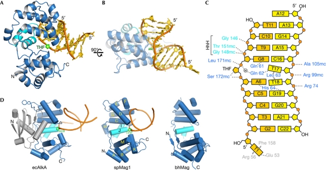
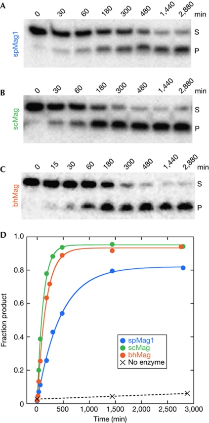
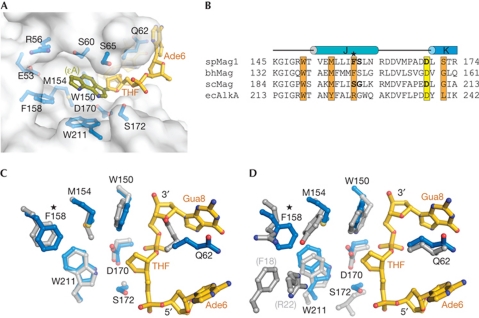
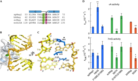
DNA glycosylases specialized for the repair of alkylation damage must identify, with fine specificity, a diverse array of subtle modifications within DNA. The current mechanism involves damage sensing through interrogation of the DNA duplex, followed by more specific recognition of the target base inside the active site pocket. To better understand the physical basis for alkylpurine detection, we determined the crystal structure of Schizosaccharomyces pombe Mag1 (spMag1) in complex with DNA and performed a mutational analysis of spMag1 and the close homologue from Saccharomyces cerevisiae (scMag). Despite strong homology, spMag1 and scMag differ in substrate specificity and cellular alkylation sensitivity, although the enzymological basis for their functional differences is unknown. We show that Mag preference for 1,N(6)-ethenoadenine (ɛA) is influenced by a minor groove-interrogating residue more than the composition of the nucleobase-binding pocket. Exchanging this residue between Mag proteins swapped their ɛA activities, providing evidence that residues outside the extrahelical base-binding pocket have a role in identification of a particular modification in addition to sensing damage.


Conflict of interest statement

The authors declare that they have no conflict of interest.

Figures

Figure 1
Figure 1
Structures of alkylated bases relevant to this study.
Figure 2
Figure 2
The spMag1–DNA complex crystal structure. (A,B) Orthogonal views of spMag1 (blue ribbons) bound to DNA (gold) containing a tetrahydrofuran (THF) abasic site analogue (green). The helix–hairpin–helix (HhH) motif is light blue. (C) Schematic of spMag1–DNA interactions, with protein residues in blue, THF–DNA strand in gold, undamaged DNA strand in yellow and phosphates depicted as orange circles. Dotted and wavy lines represent hydrogen bonds and van der Waals interactions, respectively. (D) Structural alignment of 3-methyladenine DNA glycosylases: E. coli AlkA bound to 1-azaribose (aza) DNA (ecAlkA, PDB ID 1DIZ), S. pombe Mag1 and B. halodurans Mag (bhMag, PDB ID 2H56).
Figure 3
Figure 3
1,N6-ethenoadenine (εA) excision activity of Mag orthologues. (AC) Denaturing polyacrylamide gels showing the disappearance of εA-containing DNA substrate (S) and appearance of alkaline-cleaved abasic-DNA product (P) as a function of time after addition of spMag1 (A), scMag (B) and bhMag (C). (D) Quantification of the panels shown in A–C. Blue, spMag1; green, scMag; orange, bhMag. Rate constants calculated from the single-exponential fits to the data are shown in supplementary Table S3 online.
Figure 4
Figure 4
SpMag1 nucleobase-binding pocket. (A) The base-binding pocket is shown as a transparent van der Waals surface. Protein side chains likely to contact an extrahelical base are shown in blue and the tetrahydrofuran (THF) abasic site and the two flanking nucleotides are shown in gold. The 1,N6-ethenoadenine (εA) base (olive) is modelled within the pocket from coordinates of εA–DNA bound to human alkyladenine DNA glycosylase (AAG; PDB ID 1F4R). (B) Sequence alignment of spMag1, bhMag and ecAlkA structures with scMag sequence. Residues predicted to contact the substrate base are highlighted orange, and the conserved helix–hairpin–helix (HhH) aspartic acid is yellow. The nonconserved Phe 158 is marked with a star. (C,D) Superposition of spMag1 (blue side chains, gold DNA) onto bhMag (C) and ecAlkA (D, grey).
Figure 5
Figure 5
DNA interrogation by spMag1. (A) Structure-based sequence alignment of the αC–αD loops from Mag and AlkA enzymes. Plug/wedge residues are highlighted green/light green, and the new His 64 contact in spMag1 is magenta. His64 is labeled with a star in panels A, B, and C. (B) The spMag1 αC–αD loop is shown in blue within a transparent molecular surface and intercalating residues are rendered as sticks. The damaged DNA strand is coloured gold and tetrahydrofuran (THF) green. (C) Close-up of spMag1–DNA contacts at the lesion. (D) Single-turnover rates (kcat) of 1,N6-ethenoadenine (εA; top) and 7mG (N7-methylguanine; bottom) excision are plotted for wild-type and mutant spMag1 (blue), scMag (green) and bhMag (orange). Values are shown in supplementary Table S3 online and raw data are shown in supplementary Fig S6 online. Error bars represent the standard deviation (n=3). Asterisks denote P<0.05 (*) and P<0.002 (**).

Similar articles

Cited by

References

    1. Alseth I, Osman F, Korvald H, Tsaneva I, Whitby MC, Seeberg E, Bjoras M (2005) Biochemical characterization and DNA repair pathway interactions of Mag1-mediated base excision repair in Schizosaccharomyces pombe. Nucleic Acids Res 33: 1123–1131 - PMC - PubMed
    1. Banerjee A, Santos WL, Verdine GL (2006) Structure of a DNA glycosylase searching for lesions. Science 311: 1153–1157 - PubMed
    1. Barnes DE, Lindahl T (2004) Repair and genetic consequences of endogenous DNA base damage in mammalian cells. Annu Rev Genet 38: 445–476 - PubMed
    1. Bowman BR, Lee S, Wang S, Verdine GL (2010) Structure of Escherichia coli AlkA in complex with undamaged DNA. J Biol Chem 285: 35783–35791 - PMC - PubMed
    1. Chen J, Derfler B, Samson L (1990) Saccharomyces cerevisiae 3-methyladenine DNA glycosylase has homology to the AlkA glycosylase of E. coli and is induced in response to DNA alkylation damage. EMBO J 9: 4569–4575 - PMC - PubMed

Publication types

MeSH terms

Associated data