. 2011 Dec;32(11):2126-31.
doi: 10.3174/ajnr.A2707. Epub 2011 Sep 29.

Erdheim-Chester disease of the central nervous system: new manifestations of a rare disease

Affiliations

Erdheim-Chester disease of the central nervous system: new manifestations of a rare disease

P Sedrak et al. AJNR Am J Neuroradiol. 2011 Dec.

Abstract

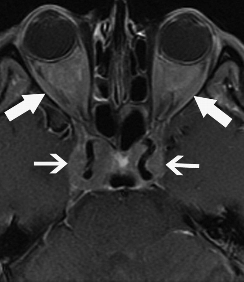
Background and purpose: ECD is a rare non-Langerhans-cell histiocytosis, which can involve the CNS; therefore, CNS imaging findings have been described in only a small number of patients. To gain additional insight into the CNS manifestations of ECD, we reviewed the findings on imaging of the brain, head and neck, and spine in patients with ECD who presented to our institution. Here, we illustrate manifestations that have not, to our knowledge, been previously described.

Materials and methods: CT, MR imaging, and PET/CT studies of the brain, maxillofacial region, and spine were reviewed in 11 patients with ECD.

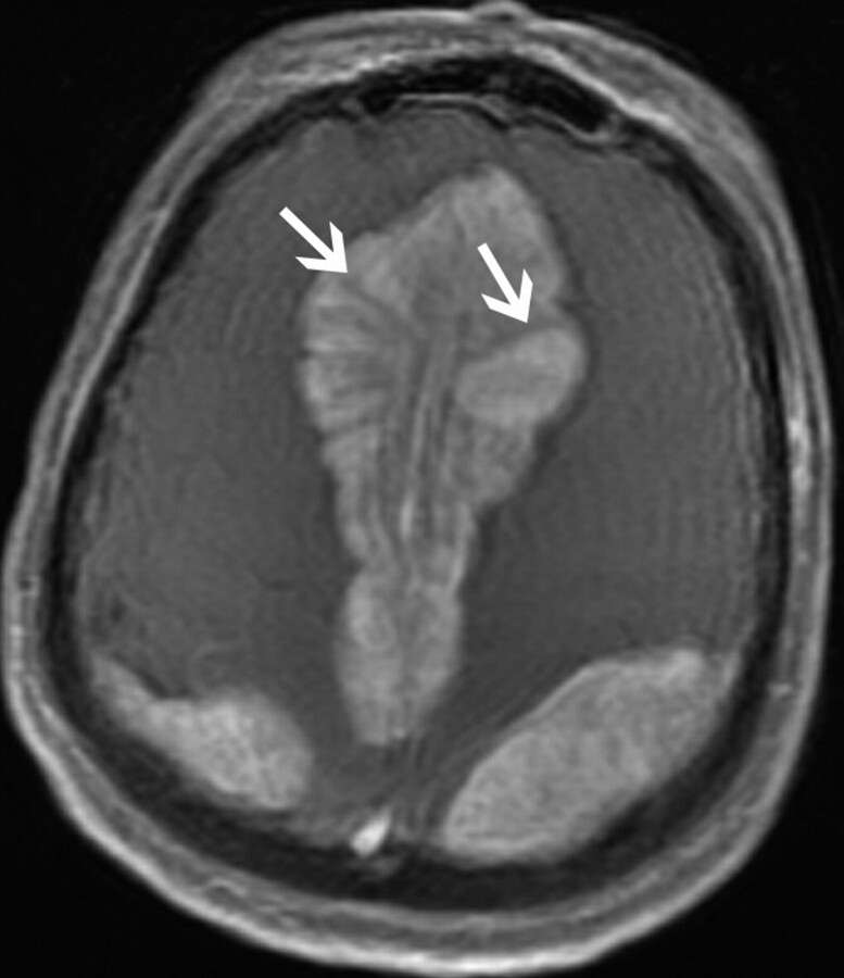
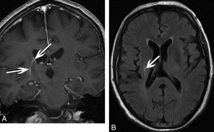
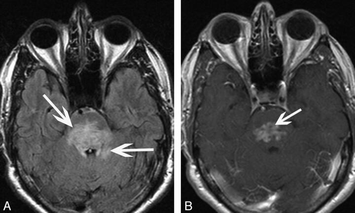
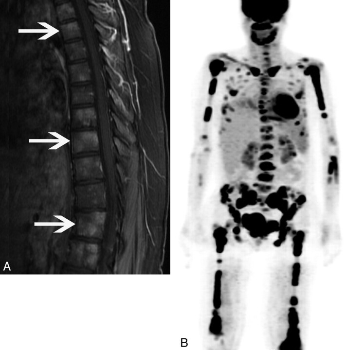
Results: Four new manifestations of ECD were present, including the following: a stellate appearance of intracranial extra-axial lesions, ependymal enhancement along the lateral ventricle with deep linear extension to the lentiform nucleus, irregular enhancement in the pons, and diffuse involvement of the vertebral column on PET/CT.

Conclusions: ECD has a variety of imaging appearances in the CNS, including new manifestations described herein. Neuroradiologists should be aware of these manifestations to avoid mistaking them for other disease processes.


Figures

Fig 1.
Fig 1.
A 48-year-old woman (patient 10) who presented with fatigue and mental status change. Axial T1-weighted postgadolinium image of a bilateral parasagittal extra-axial mass demonstrates a stellate appearance, without enhancement of the spicules (arrows).
Fig 2.
Fig 2.
A 64-year-old man (patient 11) who presented with diabetes insipidus. A, Coronal T1-weighted postgadolinium image of an ependymal enhancing focus extending inferiorly toward the right lentiform nucleus (arrows). B, Axial FLAIR image demonstrates corresponding FLAIR signal-intensity hyperintensity (arrow).
Fig 3.
Fig 3.
A 62-year-old man (patient 6) who presented with ataxia. A, Axial FLAIR image demonstrates hyperintensity in the pons with extension into the adjacent cerebellum (arrows). B, Axial T1-weighted postgadolinium image shows irregular heterogeneous enhancement oriented transversely along the pons (arrow).
Fig 4.
Fig 4.
A 49-year-old man (patient 8) who presented with diabetes insipidus and fatigue. Axial T1-weighted postgadolinium image shows sellar and cavernous sinus involvement surrounding the cavernous carotid arteries (small arrows). Intra- and extraconal orbital involvement is also present (large arrows).
Fig 5.
Fig 5.
A 37-year-old woman (patient 1) who presented with gait instability. A, Sagittal T1-weighted postgadolinium image shows heterogeneously enhancing foci throughout the vertebral column (arrows). B, 3D PET further demonstrates diffuse FDG avidity in the spine, appendicular skeleton, and mandible.
Fig 6.
Fig 6.
A 49-year-old man (patient 8, also described in Fig 4) with retro-orbital lesions. A, The orbital soft tissue is cellular with infiltration by predominance of foamy histiocytes (arrow) (hematoxylin-eosin, original magnification ×200). B, The cells are positive for CD68 (KP-1), consistent with histocytic origin (arrows) (CD68 immunohistochemical study, original magnification ×200). The cellular infiltrate is notably negative for CD1a on the CD1a immunohistochemical study (not shown).

References

    1. Veyssier-Belot C, Cacoub P, Caparros-Lefebvre D, et al. . Erdheim-Chester disease: clinical and radiologic characteristics of 59 cases. Medicine 1996;75:157–69 - PubMed
    1. Martinez R. Erdheim-Chester disease: MR of intraaxial and extraaxial brain stem lesions. AJNR Am J Neuroradiol 1995;16:1787–90 - PMC - PubMed
    1. Bohlega S, Alwatban J, Tulbah A, et al. . Cerebral manifestation of Erdheim-Chester disease: clinical and radiologic findings. Neurology 1997;49:1702–05 - PubMed
    1. Gottlieb R, Chen A. MR findings of Erdheim-Chester disease. J Comput Assist Tomogr 2002;26:257–61 - PubMed
    1. Drier A, Haroche J, Savatovsky J, et al. . Cerebral, facial, and orbital involvement in Erdheim-Chester disease: CT and MR imaging findings. Radiology 2010;255:586–94 - PubMed