. 2011 Dec;301(6):G1052-8.
doi: 10.1152/ajpgi.00015.2011. Epub 2011 Sep 29.

Effect of synthetic cationic protein on mechanoexcitability of vagal afferent nerve subtypes in guinea pig esophagus

Affiliations

Effect of synthetic cationic protein on mechanoexcitability of vagal afferent nerve subtypes in guinea pig esophagus

Shaoyong Yu et al. Am J Physiol Gastrointest Liver Physiol. 2011 Dec.

Abstract

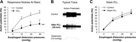
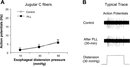
Eosinophilic esophagitis is characterized by increased infiltration and degranulation of eosinophils in the esophagus. Whether eosinophil-derived cationic proteins regulate esophageal sensory nerve function is still unknown. Using synthetic cationic protein to investigate such effect, we performed extracellular recordings from vagal nodose or jugular neurons in ex vivo esophageal-vagal preparations with intact nerve endings in the esophagus. Nerve excitabilities were determined by comparing action potentials evoked by esophageal distensions before and after perfusion of synthetic cationic protein poly-L-lysine (PLL) with or without pretreatment with poly-L-glutamic acid (PLGA), which neutralized cationic charges of PLL. Perfusion with PLL did not evoke action potentials in esophageal nodose C fibers but increased their responses to esophageal distension. This potentiation effect lasted for 30 min after washing out of PLL. Pretreatment with PLGA significantly inhibited PLL-induced mechanohyperexcitability of esophageal nodose C fibers. In esophageal nodose Aδ fibers, perfusion with PLL did not evoke action potentials. In contrast to nodose C fibers, both the spontaneous discharges and the responses to esophageal distension in nodose Aδ fibers were decreased by perfusion with PLL, which can be restored after washing out PLL for 30-60 min. Pretreatment with PLGA attenuated PLL-induced decrease in spontaneous discharge and mechanoexcitability of esophageal nodose Aδ fibers. In esophageal jugular C fibers, PLL neither evoked action potentials nor changed their responses to esophageal distension. Collectively, these data demonstrated that synthetic cationic protein did not evoke action potential discharges of esophageal vagal afferents but had distinctive sensitization effects on their responses to esophageal distension.


Figures

Fig. 1.
Fig. 1.
Nodose C fiber in response to synthetic cationic protein poly-l-lysine (PLL). A: peaks of action potential discharges evoked by esophageal distensions significantly increased from 2.75 ± 0.61, 6.15 ± 1.07, and 11.2 ± 1.31 Hz (control) to 4.8 ± 1.35, 13.2 ± 1.67, and 19.5 ± 1.91 Hz (after PLL) at distension pressures of 10, 30, and 60 mmHg for 20 s, respectively (**P < 0.05, n = 20). B: typical trace of nodose C fiber's responses (action potentials) to esophageal distension before (control) and after PLL perfusion. C: after washout of PLL for 30 min, the peaks of action potential discharges evoked by esophageal distensions remained significantly greater than control at 5.3 ± 1.1, 9.4 ± 1.4, and 17.4 ± 1.8 Hz (compared with control of 2.3 ± 0.3, 4.8 ± 0.4, and 8.8 ± 0.9 Hz, at distension pressures of 10, 30, and 60 mmHg for 20 s, respectively, **P < 0.05, n = 10).
Fig. 2.
Fig. 2.
Nodose C fiber in response to synthetic cationic protein PLL after pretreatment with poly-l-glutamic acid (PLGA). Compared with control, PLL perfusion did not significantly change the action potential discharges evoked by esophageal distension after perfusion with and continually in the presence of PLGA (control: 3.1 ± 0.9, 7.3 ± 1.7, and 11.7 ± 2.0 Hz vs. PLGA+PLL: 3.8 ± 0.9, 7.8 ± 1.6, and 12.5 ± 2.0 Hz at distension pressures of 10, 30, and 60 mmHg for 20 s, respectively, P > 0.05, n = 10).
Fig. 3.
Fig. 3.
Nodose Aδ fiber in response to synthetic cationic protein PLL. A: in nodose Aδ fibers, PLL perfusion (50 μg/0.1 ml in Krebs' solution, 30 min) did not evoke action potential discharges, but the spontaneous action potential discharges (showing at distension pressure of 0 mmHg) decreased from 5.2 ± 0.5 Hz before to 2.2 ± 1.0 Hz after PLL perfusion (P < 0.05, n = 8). The action potential discharges evoked by esophageal distensions decreased significantly from 24.0 ± 4.1, 30.3 ± 3.9, and 34.6 ± 4.2 Hz (control) to 7.4 ± 4.8, 12.6 ± 5.9, and 14.9 ± 7.0 Hz (after PLL) at distension pressures of 10, 30, and 60 mmHg for 20 s, respectively (P < 0.05, n = 8). B: typical trace of nodose Aδ fiber's response (action potentials) to esophageal distension before and after PLL perfusion. C: after PLL was washed out for 30–60 min, the peaks of action potential discharges evoked by esophageal distensions were restored to 20.3 ± 1.8, 27.5 ± 2.6, and 33.3 ± 4.5 Hz at distension pressures of 10, 30, and 60 mmHg for 20 s, respectively (vs. control, P > 0.05, n = 4).
Fig. 4.
Fig. 4.
Nodose Aδ fiber in response to synthetic cationic protein PLL after pretreatment with PLGA. Compared with control, PLL did not significantly changed the action potential discharges evoked by esophageal distension after perfusion with and in the continued presence of PLGA (control: 20.4 ± 3.0, 27.7 ± 2.2, and 28.9 ± 3.0 Hz vs. PLL+PLGA: 16.7 ± 0.9, 22.6 ± 2.6, and 23.3 ± 2.8 Hz at distension pressures of 10, 30, and 60 mmHg for 20 s, respectively, P > 0.05, except at pressure of 60 mmHg, n = 8).
Fig. 5.
Fig. 5.
Jugular C fiber in response to synthetic cationic protein PLL. A: peaks of action potential discharges evoked by esophageal distensions did not change before and after PLL perfusion (1.4 ± 0.2, 3.0 ± 0.5, and 6.2 ± 0.8 vs. 1.6 ± 0.3, 3.3 ± 0.6, and 5.7 ± 0.9 Hz at distension pressures of 10, 30, and 60 mmHg for 20 s, respectively, P > 0.05, n = 10). B: typical trace of jugular C fiber's responses (action potentials) to esophageal distension before and after PLL perfusion.

References

    1. Achem SR, Almansa C, Krishna M, Heckman MG, Wolfsen HC, Talley NJ, DeVault KR. Oesophageal eosinophilic infiltration in patients with noncardiac chest pain. Aliment Pharmacol Ther 33: 1194–1201, 2011 - PubMed
    1. Allende DS, Yerian LM. Diagnosing gastroesophageal reflux disease: the pathologist's perspective. Adv Anat Pathol 16: 161–165, 2009Review - PubMed
    1. Blackshaw LA, Brookes SJ, Grundy D, Schemann M. Sensory transmission in the gastrointestinal tract. Neurogastroenterol Motil 19, Suppl 1: 1–19, 2007 - PubMed
    1. Coyle AJ, Ackerman SJ, Irvin CG. Cationic proteins induce airway hyperresponsiveness dependent on charge interactions. Am Rev Respir Dis 147: 896–900, 1993 - PubMed
    1. Durcan N, Costello RW, McLean WG, Blusztajn J, Madziar B, Fenech AG, Hall IP, Gleich GJ, McGarvey L, Walsh MT. Eosinophil-mediated cholinergic nerve remodeling. Am J Respir Cell Mol Biol 34: 775–786, 2006 - PubMed

Publication types

MeSH terms

Substances

LinkOut - more resources