Comparative Study
. 2011 Oct 31;52(11):8505-13.
doi: 10.1167/iovs.11-8194.

αVβ6 integrin promotes corneal wound healing

Affiliations
Comparative Study

αVβ6 integrin promotes corneal wound healing

José Tomás Blanco-Mezquita et al. Invest Ophthalmol Vis Sci. .

Abstract

Purpose: To appreciate the role of the integrin αvβ6 in corneal wound repair, corneal debridement and keratectomy in β6 knockout (β6(-/-)) mice were examined.

Methods: Either a 2-mm debridement or keratectomy was made in 129SVE wild type mice (WT) and β6(-/-) mice and allowed to heal for up to 4 months. The pattern of corneal restoration was studied "in vivo" by slit lamp and in tissue sections by means of both light and electron microscopy. In addition, αvβ6, α6β4, laminin, and fibronectin were evaluated by indirect immunofluorescence microscopy and/or Western blot analysis.

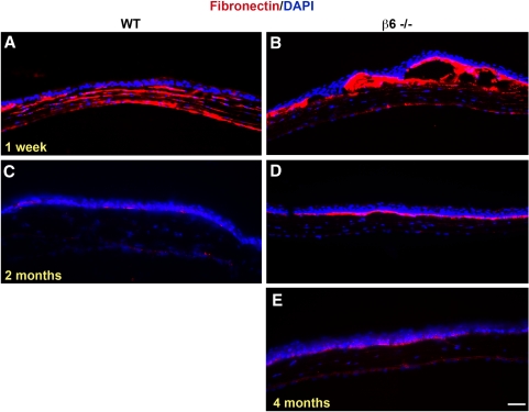
Results: αvβ6 expression was upregulated in migrating corneal epithelium after a keratectomy. Healing rates were unaffected in debridement wounds, but were significantly slowed in keratectomy wounds. Most dramatically, mice lacking αvβ6 had a severe defect in basement membrane zone (BMZ) regeneration. Levels of laminin were greatly reduced and no BMZ reformation was observed in transmission electron microscopy (TEM). In addition, hemidesmosome reformation was also impaired in the β6(-/-) mice. Analysis of the hemidesmosome component α6β4 indicated that normal amounts of this integrin were synthesized, suggesting that the defect was in reassembly of the hemidesmosomes. Finally, fibronectin persisted in the BMZ for as long as 4 months after keratectomy in the β6(-/-) mice.

Conclusions: It is hypothesized that the lack of αvβ6 leads to reduced laminin production during wound repair. This lack of laminin prevents reassembly of the BMZ and mature hemidesmosomes after keratectomy in β6(-/-) mice.


Figures

Figure 1.
Figure 1.
Immunolocalization of αvβ6 (green) in WT mouse corneas: (A) unwounded, (B) 24 hours, (C) 48 hours, (D) 4 days, and (E) 4 weeks after keratectomy. (C, inset) WT mouse cornea 48 hours after debridement. In (F) and (G) whole mount corneas were examined by confocal microscopy 48 hours after keratectomy. (G) A 3-D software reconstruction (Image Pro Plus v.7; Media Cybernetics). Note the abrupt increase of expression in the basal cells migrating to cover the wound area. Blue in (G), DAPI, a marker of cell nuclei. Arrows denote the original wound edge. Bar, 50 μm.
Figure 2.
Figure 2.
Western blot analysis of αvβ6 in WT mouse corneas after a keratectomy wound. (A) Representative Western blot images documenting the increased expression of αvβ6, and the corresponding stain (Ponceau S; Sigma) of the blot to document equal loading of samples (region from 40 to 70 kDa). Experiments were repeated three times using at least six mice per time point. (B) Density of the bands, quantitated and plotted.
Figure 3.
Figure 3.
Graphical documentation of healing rates of WT and β6−/− mice after 2-mm debridement or keratectomy. At least six mice were used per time point. ** P < 0.01, *** P < 0.001.
Figure 4.
Figure 4.
Morphologic analysis of WT (A, C, E, G) and β6−/− (B, D, F, H) in (A, B) unwounded corneas, (C, D) 4 days, (E, F) 2 weeks, and (G, H) 4 weeks post keratectomy. Note that epithelium is loosely adherent in β6−/− after wounding. Bar, 100 μm. (I) Slit lamp image (magnification, ×40) of β6−/− cornea 1 week after keratectomy. Note the blister-like structure extends across the cornea (arrows).
Figure 5.
Figure 5.
Transmission electron micrographs of WT (A, C, E) and β6−/− (B, D, F) mouse corneas 1 month after debridement (A, B), and 1 (C, D) and 4 months (E, F) post keratectomy. Note the complete absence of BMZ and the greatly reduced number of electron dense hemidesmosomes 1 month after keratectomy in the β6−/− mice. Also note the epithelial projections into the stroma that may help anchor the epithelium. Arrows indicate location of BMZ. Bar, 500 nm.
Figure 6.
Figure 6.
Immunolocalization of laminin (A–I, red) and α6β4 (A1–I1, green) in WT (A–D, A1–D1) and β6−/− (E–I, E1–I1) mice. Proteins were localized in unwounded corneas (A, A1, E, E1) and 4 days (B, B1, F, F1), 1 week (C, C1, G, G1), 2 weeks (H, H1), 2 months (D, D1) and 4 months (I, I1) post keratectomy. Note that laminin and α6β4 were analyzed on single sections that are shown separately for ease of viewing. DAPI, a marker of all cell nuclei, blue. Bar, 50 μm. At least four corneas per time point were analyzed.
Figure 7.
Figure 7.
Representative Western blot images documenting the increased expression of laminin (1A) and α6β4 (2A) after keratectomy wounds. Stain (Ponceau S; Sigma) of the blots was used to document equal loading of samples (data not shown). Experiments were repeated two or three times using at least six mice per time point. The density of the bands was quantitated and fold enhancement was plotted (1B, 2B). Note: Only epithelium was included in these samples.
Figure 8.
Figure 8.
Immunolocalization of fibronectin (red) in WT (A, C) and β6−/− (B, D, E) mice: (A, B) 1 week, (C, D) 2 and (E) 4 months post keratectomy. Note that no fibronectin was present in unwounded corneas, or beyond 2 weeks after wounding in WT cornea. DAPI, a marker of all cell nuclei, blue. Bar, 50 μm.

References

    1. Ruoslahti E, Pierschbacher MD. New perspectives in cell adhesion: RGD and integrins. Science. 1987;238:491–497 - PubMed
    1. Hynes RO. Integrins: a family of cell surface receptors. Cell. 1987;48:549–554 - PubMed
    1. Clark EA, Brugge JS. Integrins and signal transduction pathways: the road taken. Science. 1995;268:233–239 - PubMed
    1. Breuss JM, Gillett N, Lu L, Sheppard D, Pytela R. Restricted distribution of integrin beta 6 mRNA in primate epithelial tissues. J Histochem Cytochem. 1993;41:1521–1527 - PubMed
    1. Breuss JM, Gallo J, DeLisser HM, et al. Expression of the beta 6 integrin subunit in development, neoplasia and tissue repair suggests a role in epithelial remodeling. J Cell Sci. 1995;108:2241–2251 - PubMed

Publication types

MeSH terms