. 2012 Jan;112(1):204-11.
doi: 10.1152/japplphysiol.00953.2011. Epub 2011 Sep 29.

Microparticle enlargement and altered surface proteins after air decompression are associated with inflammatory vascular injuries

Affiliations

Microparticle enlargement and altered surface proteins after air decompression are associated with inflammatory vascular injuries

Ming Yang et al. J Appl Physiol (1985). 2012 Jan.

Abstract

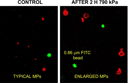
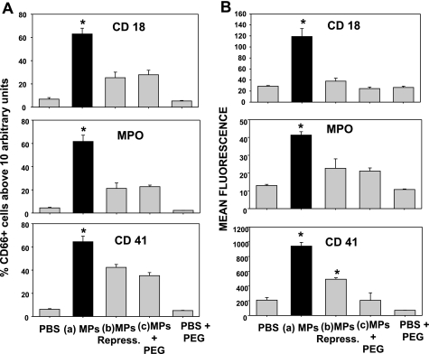
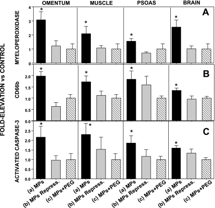
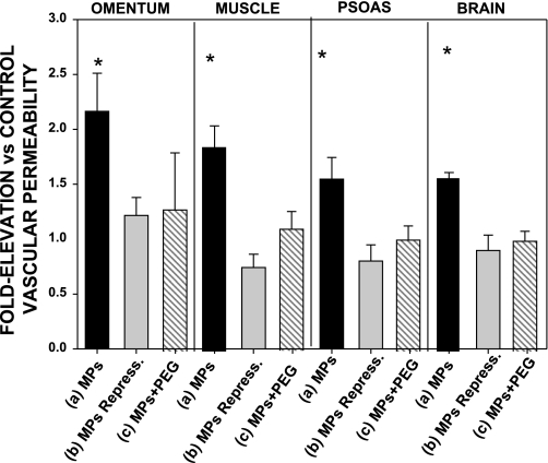
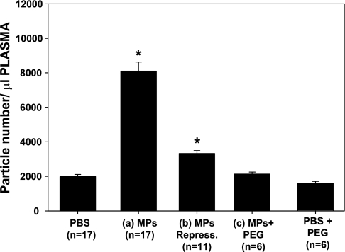
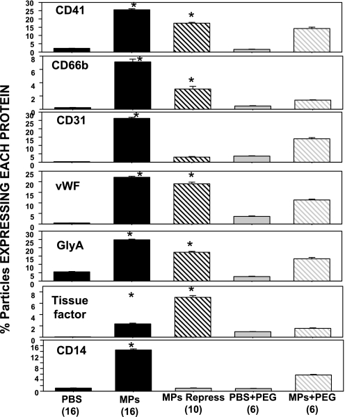
Studies in a murine model have shown that decompression stress triggers a progressive elevation in the number of circulating annexin V-coated microparticles derived from leukocytes, erythrocytes, platelets, and endothelial cells. We noted that some particles appeared to be larger than anticipated, and size continued to increase for ≥24 h postdecompression. These observations led to the hypothesis that inert gas bubbles caused the enlargement and particle size could be reduced by hydrostatic pressure. After demonstrating pressure-induced particle size reduction, we hypothesized that annexin V-positive particle changes associated with decompression contributed to their proinflammatory potential. Intravenous injection of naive mice with particles isolated from decompressed mice, but not control mice, caused intravascular neutrophil activation; perivascular neutrophil sequestration and tissue injuries were documented as elevations of vascular permeability and activated caspase-3. These changes were not observed if mice were injected with particles that had been subjected to hydrostatic recompression or particles that had been emulsified by incubation with polyethylene glycol telomere B surfactant. Hydrostatic pressure and surfactant incubation also altered the pattern of proteins expressed on the surface of particles. We conclude that proinflammatory events and vascular damage are due to enlargement of annexin V-coated particles and/or changes in surface marker protein pattern associated with provocative decompression. Injection of annexin V-coated particles from decompressed mice will recapitulate the pathophysiological vascular changes observed following decompression stress.


Figures

Fig. 1.
Fig. 1.
Confocal microscopy images of annexin V-positive microparticles (MPs). Image frames include 0.86-μm-diameter green fluorescent (FITC) beads. Samples prepared from decompressed mice exhibited numerous annexin V-positive particles that appeared to have diameters >1 μm.
Fig. 2.
Fig. 2.
Flow cytometric analysis of size distribution of blood-borne annexin V-positive particles postdecompression. Blood from control mice and mice exposed to 790 kPa air pressure for 2 h was fixed immediately after it was withdrawn to prevent MP enlargement. Analysis was based on comparison of annexin V-positive particles with fluorescent beads added to each suspension prior to analysis. Values are means ± SE; n = 3–4 in all groups. *P < 0.05 vs. control.
Fig. 3.
Fig. 3.
Neutrophil activation due to MP injections. Neutrophils were identified by CD66b staining, and coexpression of CD18, myeloperoxidase (MPO), and CD41 was assessed by flow cytometry. A: percentage of CD66b-positive cells expressing a geometric mean fluorescence value ≥10 arbitrary fluorescence units for each surface marker. B: CD66b-positive cell geometric mean fluorescence for each marker. PBS, injections of sterile saline only; MPs, annexin V-positive particles isolated from decompressed mice and left on the benchtop for 1 h at ambient pressure; MPs Repress, MPs subjected to hydrostatic recompression for 1 h; PEG + MPs, MPs incubated with polyethylene glycol telomere B (PEG); PBS + PEG, injection with PBS containing PEG but no MPs. Values are means ± SE; n = 4–10 per group. *P < 0.05 vs. control.
Fig. 4.
Fig. 4.
MPO, CD66b, and activated caspase-3 in tissue homogenates. Tissues were isolated, enriched for endothelium, and analyzed. Western blot band densities are expressed relative to band density of tissue from control animals (injected with PBS) run on the same blot (therefore, control value = 1.0). Mice were injected with solutions as described in Fig. 3 legend. Data for mice injected with PBS + PEG were virtually identical to data for control mice injected with PBS (data not shown). Values are means ± SE; n = 4–10 for each group. *P < 0.05 vs. control.
Fig. 5.
Fig. 5.
Rhodamine-labeled dextran uptake, measured as leakage of lysine-fixable rhodamine-conjugated 2 × 10⁶ Da dextran, in mice injected with solutions as described in Fig. 3 legend. Values reflect ratio of fluorescence/mg protein in homogenates from injected mice to that in homogenates from control mice injected with the same lot of rhodamine-dextran and run on the same day. Data for mice injected with PBS + PEG were virtually identical to data for control mice injected with PBS (data not shown). Values are means ± SE; n = 4–10 for each group. *P < 0.05 vs. control.
Fig. 6.
Fig. 6.
Circulating annexin V-positive particles in mice killed 1 h after injections with various preparations as described in Fig. 3 legend. Values are means ± SE for number of animals shown for each group (n). *P < 0.05 vs. control (PBS). Values for MPs Repress and MPs are significantly different from each other.
Fig. 7.
Fig. 7.
Proteins expressed on the surface of MPs: fractions of MPs expressing the platelet-specific CD41, the neutrophil protein CD66b, CD31 (platelet endothelial cell adhesion molecule), von Willebrand factor (vWF), the erythrocyte-specific protein glycophorin A (GlyA), tissue factor, and the common leukocyte antigen CD14. Values are means ± SE for number of animals shown (in parentheses) for each group. *P < 0.05 vs. control (PBS).

References

    1. Altman PL, Dittmer DS. Biology Databook. Washington, DC: FASEB, 1964, p. 264
    1. Brubakk AO, Duplancic D, Valic Z, Palada I, Obad A, Bakovic D, Wisloff U, Dujic Z. A single air dive reduces arterial endothelial function. J Physiol 566: 901–906, 2005 - PMC - PubMed
    1. Connor DE, Exner T, Ma DD, Joseph JE. The majority of circulating platelet-derived microparticles fail to bind annexin V, lack phospholipid-dependent procoagulant activity and demonstrate greater expression of glycoprotein Ib. Thromb Haemost 103: 1044–1052, 2010 - PubMed
    1. Eftedal OS, Lydersen S, Brubakk AO. The relationship between venous gas bubbles and adverse effects of decompression after air dives. Undersea Hyperb Med 34: 99–105, 2007 - PubMed
    1. Enjeti AK, Lincz LF, Seldon M. Detection and measurement of microparticles: an evolving research tool for vascular biology. Semin Thromb Hemost 33: 771–779, 2007 - PubMed

Publication types