. 2011:2011:481064.
doi: 10.1155/2011/481064. Epub 2011 Sep 25.

Patient Specific Dosimetry Phantoms Using Multichannel LDDMM of the Whole Body

Affiliations

Patient Specific Dosimetry Phantoms Using Multichannel LDDMM of the Whole Body

Daniel J Tward et al. Int J Biomed Imaging. 2011.

Abstract

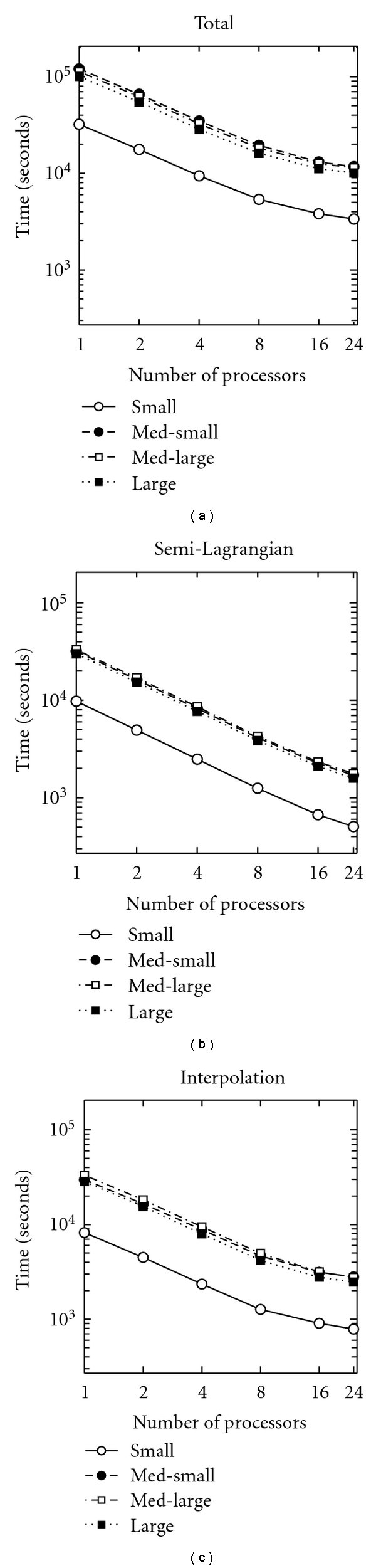
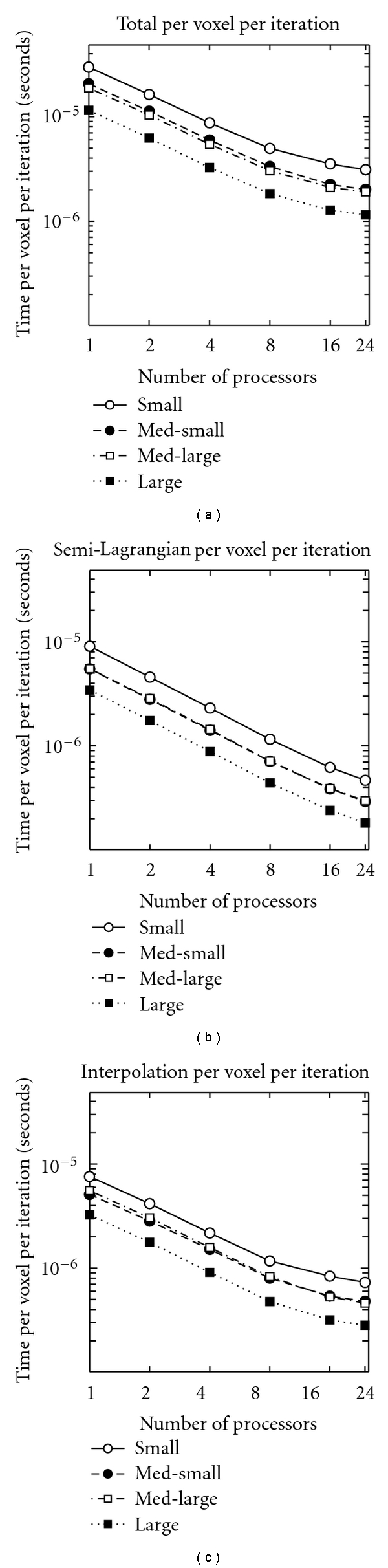
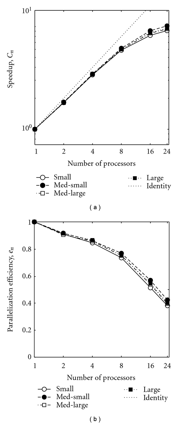
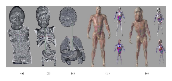
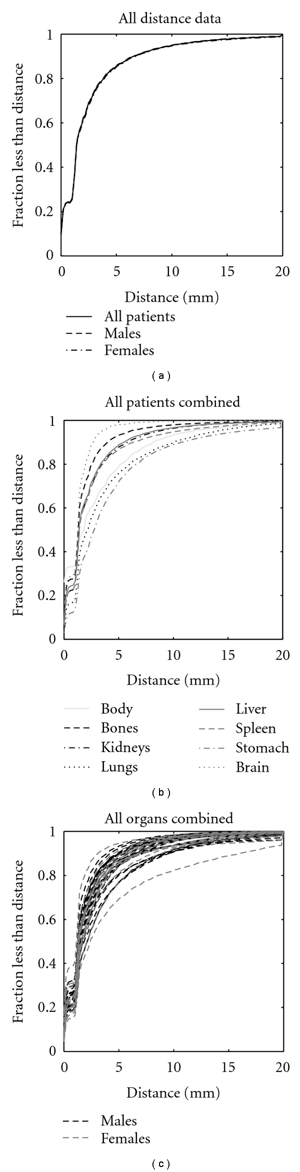
This paper describes an automated procedure for creating detailed patient-specific pediatric dosimetry phantoms from a small set of segmented organs in a child's CT scan. The algorithm involves full body mappings from adult template to pediatric images using multichannel large deformation diffeomorphic metric mapping (MC-LDDMM). The parallel implementation and performance of MC-LDDMM for this application is studied here for a sample of 4 pediatric patients, and from 1 to 24 processors. 93.84% of computation time is parallelized, and the efficiency of parallelization remains high until more than 8 processors are used. The performance of the algorithm was validated on a set of 24 male and 18 female pediatric patients. It was found to be accurate typically to within 1-2 voxels (2-4 mm) and robust across this large and variable data set.


Figures

Figure 1
Figure 1
An example of how the standard MC-LDDMM algorithm fails for full body mapping. (a) axial, (b) coronal, and (c) sagittal images of a deformed adult template. Notice that the abdominal organs have been catastrophically shrunk causing distortions in nearby neck and thoracic structures and that details in the face and skull have been lost.
Figure 2
Figure 2
The robust sequence of transformations leading to the final mapping. Top row: sagittal slice, middle row: coronal slice, bottom row: axial slice. (a) Initial placement, (b) after affine registration, (c) after LDDMM landmark, and (d)–(g) after 1–4 iterations of MC-LDDMM.
Figure 3
Figure 3
Time spent on computations for the four patients examined, plotted on a log-log axis. (a) Total time, (b) time in semi-Lagrangian advection, (c) time in image interpolation. Note that in (a) med-small takes the longest, followed by med-large, large, and small. In (b) and (c), the order of the first two is reversed.
Figure 4
Figure 4
Time spent on computations, per image voxel per gradient descent iteration, for the four patients examined, plotted on a log-log axis. (a) Total time, (b) time in semi-Lagrangian advection, and (c) time in image interpolation. Note that in (a) small takes the longest, followed by med-small, med-large, and large. In (b) (for all processors) and (c) (from 1 to 8 processors), the order of the middle two is reversed.
Figure 5
Figure 5
(a) Speedup due to parallelization (log scale) and (b) efficiency of parallelization (semilog scale), for the four patients examined. With the exception of “small” being uniformly the lowest, the order of the other varies as number of processors increases, and differences between each curve are quite small.
Figure 6
Figure 6
Triangulated surfaces from an example deformed adult template (white) and target child (black) are of (a) body, (b) bones, and (c) other organs. Adult male XCAT phantom is shown in (d), and an example custom dosimetry phantom is shown in (e).
Figure 7
Figure 7
Cumulative distribution functions of final surface to surface distances are shown for all data and for all males and all females in (a), for individual organs with all patients combined in (b), and for individual patients with all organs combined in (c).

References

    1. Brenner DJ, Hall EJ. Computed tomography—an increasing source of radiation exposure. The New England Journal of Medicine. 2007;357(22):2277–2284. - PubMed
    1. The Alliance for Radiation Safety in Pediatric Imaging. “Image gently.”, 2009, http://www.pedrad.org/associations/5364/ig.
    1. Lee C, Lodwick D, Williams JL, Bolch WE. Hybrid computational phantoms of the 15-year male and female adolescent: applications to CT organ dosimetry for patients of variable morphometry. Medical Physics. 2008;35(6):2366–2382. - PMC - PubMed
    1. Segars WP, Mahesh M, Beck TJ, Frey EC, Tsui BMW. Realistic CT simulation using the 4D XCAT phantom. Medical Physics. 2008;35(8):3800–3808. - PMC - PubMed
    1. Segars WP, et al. Patient specific computerized phantoms to estimate dose in pediatric CT. In: Proceedings of the Medical Imaging: Physics of Medical Imaging, vol. 7258; February 2009; Lake Buena Vista, Fla, USA. p. 72580H. Proceedings of SPIE.

LinkOut - more resources