

Bacterial cellulose-hydroxyapatite nanocomposites for bone regeneration
- PMID: 21961004
- PMCID: PMC3180784
- DOI: 10.1155/2011/175362
Bacterial cellulose-hydroxyapatite nanocomposites for bone regeneration
Abstract
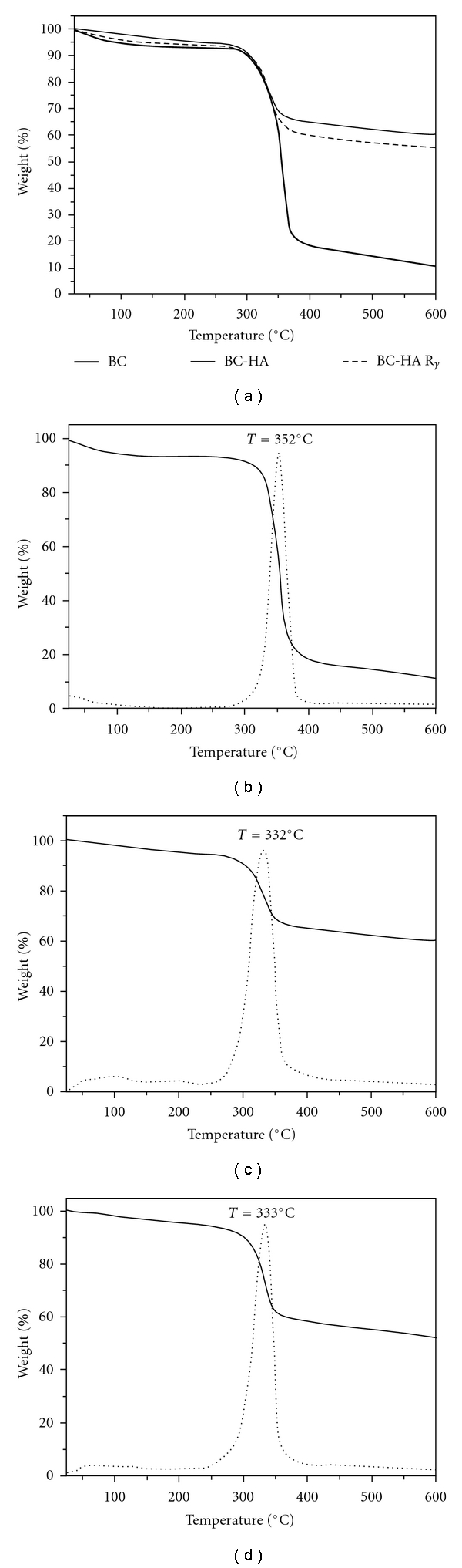
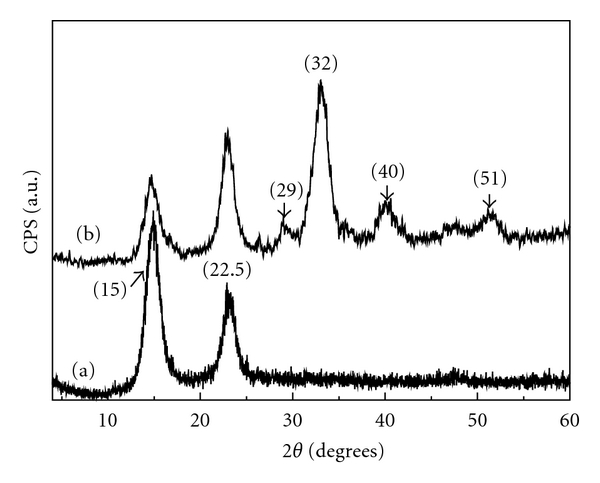
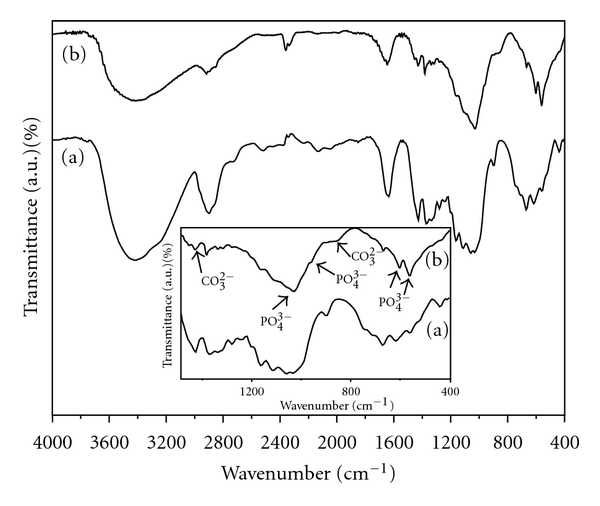
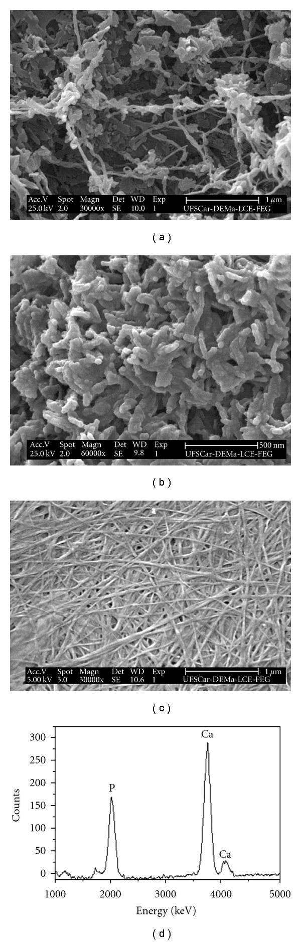
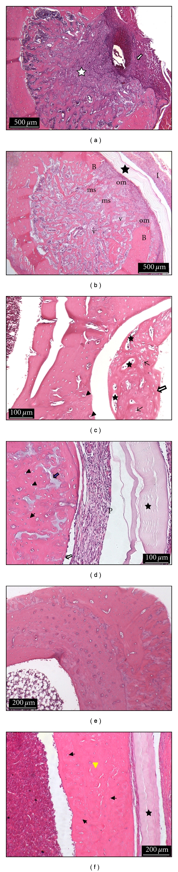
The aim of this study was to develop and to evaluate the biological properties of bacterial cellulose-hydroxyapatite (BC-HA) nanocomposite membranes for bone regeneration. Nanocomposites were prepared from bacterial cellulose membranes sequentially incubated in solutions of CaCl(2) followed by Na(2)HPO(4). BC-HA membranes were evaluated in noncritical bone defects in rat tibiae at 1, 4, and 16 weeks. Thermogravimetric analyses showed that the amount of the mineral phase was 40%-50% of the total weight. Spectroscopy, electronic microscopy/energy dispersive X-ray analyses, and X-ray diffraction showed formation of HA crystals on BC nanofibres. Low crystallinity HA crystals presented Ca/P a molar ratio of 1.5 (calcium-deficient HA), similar to physiological bone. Fourier transformed infrared spectroscopy analysis showed bands assigned to phosphate and carbonate ions. In vivo tests showed no inflammatory reaction after 1 week. After 4 weeks, defects were observed to be completely filled in by new bone tissue. The BC-HA membranes were effective for bone regeneration.
Figures
References
-
- Duskova M, Leamerova E, Sosna B, Gojis O. Guided tissue regeneration, barrier membranes and reconstruction of the cleft maxillary alveolus. Journal of Craniofacial Surgery. 2006;17(6):1153–1160. - PubMed
-
- Strietzel FP, Khongkhunthian P, Khattiya R, Patchanee P, Reichart PA. Healing pattern of bone defects covered by different membrane types-a histologic study in the porcine mandible. Journal of Biomedical Materials Research—part B. 2006;78(1):35–46. - PubMed
-
- Sculean A, Schwarz F, Chiantella GC, et al. Five-year results of a prospective, randomized, controlled study evaluating treatment of intra-bony defects with a natural bone mineral and GTR. Journal of Clinical Periodontology. 2007;34(1):72–77. - PubMed
-
- Song JH, Kim HE, Kim HW. Collagen-apatite nanocomposite membranes for guided bone regeneration. Journal of Biomedical Materials Research—part B. 2007;83(1):248–257. - PubMed
-
- Wan YZ, Huang Y, Yuan CD, et al. Biomimetic synthesis of hydroxyapatite/bacterial cellulose nanocomposites for biomedical applications. Materials Science and Engineering C. 2007;27(4):855–864.
LinkOut - more resources
Full Text Sources
Miscellaneous
