. 2011;6(9):e24349.
doi: 10.1371/journal.pone.0024349. Epub 2011 Sep 22.

Activity dependent protein degradation is critical for the formation and stability of fear memory in the amygdala

Affiliations

Activity dependent protein degradation is critical for the formation and stability of fear memory in the amygdala

Timothy J Jarome et al. PLoS One. 2011.

Abstract

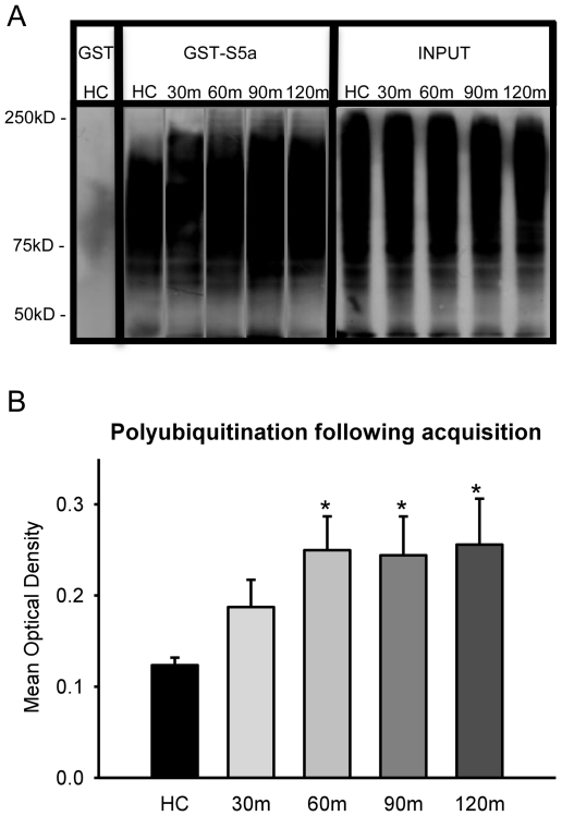
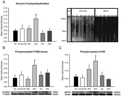
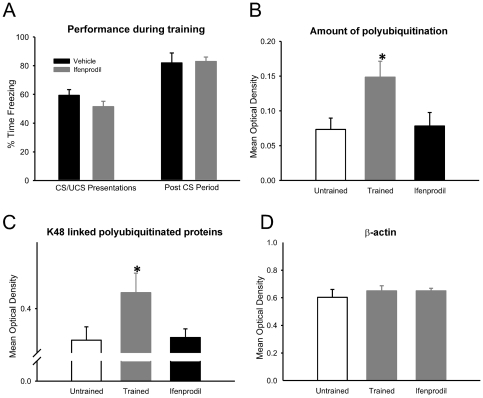
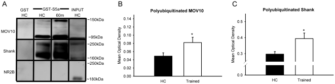
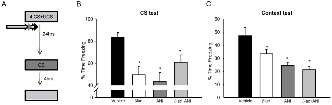
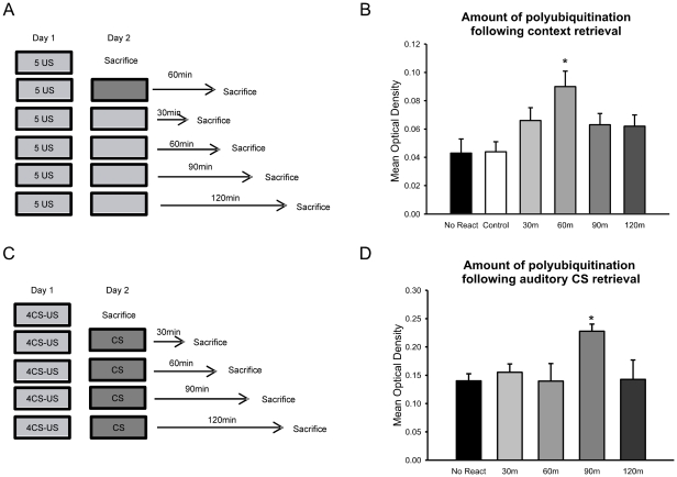
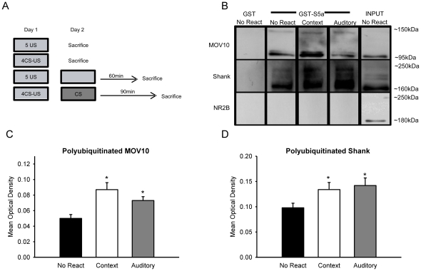
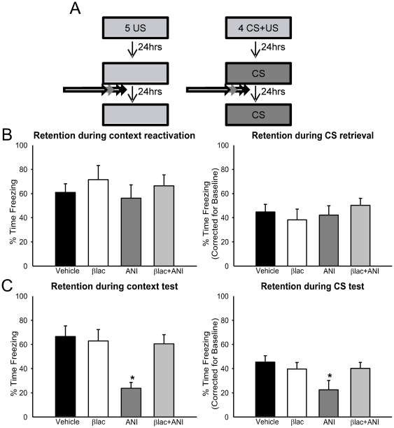
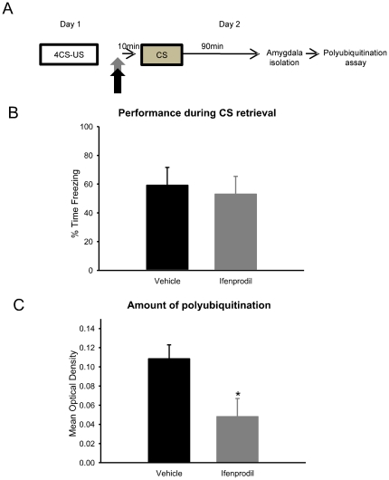
Protein degradation through the ubiquitin-proteasome system [UPS] plays a critical role in some forms of synaptic plasticity. However, its role in memory formation in the amygdala, a site critical for the formation of fear memories, currently remains unknown. Here we provide the first evidence that protein degradation through the UPS is critically engaged at amygdala synapses during memory formation and retrieval. Fear conditioning results in NMDA-dependent increases in degradation-specific polyubiquitination in the amygdala, targeting proteins involved in translational control and synaptic structure and blocking the degradation of these proteins significantly impairs long-term memory. Furthermore, retrieval of fear memory results in a second wave of NMDA-dependent polyubiquitination that targets proteins involved in translational silencing and synaptic structure and is critical for memory updating following recall. These results indicate that UPS-mediated protein degradation is a major regulator of synaptic plasticity necessary for the formation and stability of long-term memories at amygdala synapses.


Conflict of interest statement

Competing Interests: The authors have declared that no competing interests exist.

Figures

Figure 1
Figure 1. Protein degradation is increased in the amygdala following the acquisition of auditory and context fear memories.
[A] Amygdala tissue was collected in 30-min increments following fear conditioning [n = 5 per group]. Tissue was purified with GST or GST-S5a and polyubiquitinated proteins pull-downed and exposed to an antibody against ubiquitin. Input represents an aliquot of total ubiquitinated proteins. [B] There was a rapid increase in the amount of proteins targeted for UPS degradation following fear conditioning. * denotes p<.05 from homecage [HC] controls.
Figure 2
Figure 2. Increase in amygdalar protein degradation is specific to learning and mirrors protein synthesis.
Amygdala tissue was collected from naïve animals [HC, n = 8], animals exposed to either the shock [Immed SK, n = 8] or the CS [WN, n = 9], or animals that underwent fear conditioning and were sacrificed 60-min [n = 9], 6- hr [n = 9] or 24-hrs [n = 9] later and tissue was purified with GST-S5a. [A] An increase in the amount of polyubiquitinated proteins was only observed 60-min after behaviorally effective training. [B, C] Western blots with antibodies against phospho-P70S6 kinase and phospho-mTOR show that increases in protein degradation mirror increases in translational control. * denotes p<.05 from homecage [HC] controls.
Figure 3
Figure 3. Increase in amygdalar protein degradation is NMDA-dependent.
(A) Infusions of NMDA antagonist Ifenprodil (n = 8) or vehicle (n = 14) were delivered into the amygdala prior to fear conditioning, and amygdala tissue collected 60-min later and mixed with GST-S5a. Pretraining inactivation of NMDA receptors did not affect performance during training, but (B) completely abolished the increase in protein degradation. (C) Ifenprodil resulted in a significant reduction of K48-linked polyubiquitination. (D) There were no significant differences in β-actin, which was used as a loading control. * denotes p<.05 from untrained controls.
Figure 4
Figure 4. UPS targets proteins involved in translational silencing and synaptic structure in the amygdala.
Animals were trained to auditory and context fear conditioning and amygdala tissue was collected 60-min later [n = 10] and compared to naïve homecage [HC] animals [n = 10]. In all cases, crude synaptosomal membrane fractions were obtained, mixed with GST-S5a, and probed with antibodies against MOV10, Shank and NR2B [A]. The amount of polyubiquitinated MOV10 [B] and Shank [C] was increased in trained animals, suggesting potential control over protein synthesis initiation and synaptic restructuring. * denotes p<.05 from HC controls.
Figure 5
Figure 5. Protein degradation is critical for the formation of long-term fear memories.
[A] Experimental design for B-C. Animals were trained to auditory and context fear conditioning followed by infusions of βlac [n = 11], ANI [n = 12], βlac+ANI [n = 14] or vehicle [n = 10] into the amygdala. The next day, they were then tested to the auditory cue followed by the context 4-hrs later. [B, C] βlac and ANI impaired long-term memory for the auditory and context cues and simultaneous administration of both βlac+ANI did not rescue these impairments. * denotes p<.05 from Vehicle controls.
Figure 6
Figure 6. Protein degradation is increased in the amygdala following the retrieval of auditory and context fear memories.
[A] Experimental design for B. Animals were trained to context fear conditioning on Day 1. The following day, they received a 90-sec reminder to the training context and amygdala tissue was collected in 30-min increments [n = 9 per group]. A separate group of animals was placed into a novel environment and tissue collected 60-min later [n = 7]. Tissue was then purified with GST-S5a. [B] There was a rapid increase in the amount of proteins targeted for degradation, which returned to basal levels within 2-hrs. [C] Experimental design for D. Animals were trained to auditory fear conditioning on Day 1. The following day, they received a 30-sec reminder to the auditory cue and amygdala tissue was collected in 30-min increments [n = 9-10 per group]. Tissue was then purified with GST-S5a. [D] There was a delayed increase in the amount of proteins targeted for degradation, which returned to basal levels within 2-hrs. * denotes p<.05 from controls.
Figure 7
Figure 7. UPS targets proteins involved in translational silencing and synaptic structure in the amygdala following memory retrieval.
[A] Experimental design for B-D. Animals were trained to auditory or context fear conditioning on Day 1. The next day, animals were exposed to a brief retrieval and amygdala tissue collected 60-min [context, n = 10] or 90-min [auditory, n = 10] later. Two separate groups received auditory or context fear conditioning on Day 1 and were sacrificed on Day 2 without receiving retrieval [n = 6 per group]. Amygdala tissue was fractionated to obtain a crude synaptosomal membrane fraction, purified with GST-S5a, and probed with antibodies against MOV10, Shank and NR2B [B]. The amount of polyubiquitinated synaptic MOV10 [C] and Shank [D], was increased following fear memory retrieval. * denotes p<.05 from No React controls.
Figure 8
Figure 8. Protein degradation controls the destabilization of retrieved fear memories in the amygdala.
[A] Experimental design for B-C. Animals were trained to auditory or context fear conditioning on Day 1. The next day, they received a brief retrieval followed by infusions of βlac [auditory n = 13, context n = 7], ANI [auditory n = 12, context n = 7], βlac+ANI [auditory n = 12, context n = 7] or vehicle [auditory n = 13, context n = 7] into the amygdala. The next day, they were then tested to for long-term memory to their acquired cue. [B] There were no differences between groups during either context or auditory memory retrieval. [C] While βlac had no effect on either memory by itself, it rescued the memory impairments normally caused by ANI when co-infused. * denotes p<.05 from Vehicle controls.
Figure 9
Figure 9. Retrieval-induced increase in protein degradation is dependent on NMDA-receptor activity.
(A) Infusions of NMDA antagonist Ifenprodil (n = 8) or vehicle (n = 7) were delivered into the amygdala prior to fear memory retrieval, and amygdala tissue collected 90-min later and mixed with GST-S5a. (B) Pre-retrieval inactivation of NMDA receptors did not affect retention during CS retrieval, but (C) completely impaired the increase in protein degradation. * denotes p<.05 from Vehicle controls.

Similar articles

Cited by

References

    1. Davis HP, Squire LR. Protein synthesis and memory: a review. Psycho Bull. 1984;96(3):518–559. - PubMed
    1. Bailey DJ, Kim JJ, Sun W, Thompson RF, Helmstetter FJ. Acquisition of fear conditioning in rats requires the synthesis of mRNA in the amygdala. Behav Neurosci. 1999;113(2):276–282. - PubMed
    1. Parsons RG, Gafford GM, Baruch DE, Riedner BA, Helmstetter FJ. Long-term stability of fear memory depends on the synthesis of protein but not mRNA in the amygdala. Eur J Neurosci. 2006;23:1853–1859. - PMC - PubMed
    1. Schafe GE, LeDoux JE. Memory consolidation of auditory pavlovian fear conditioning requires protein synthesis and protein kinase A in the amygdala. J Neurosci. 2000;20(RC96):1–5. - PMC - PubMed
    1. Helmstetter FJ, Parsons RG, Gafford GM. Macromolecular synthesis, distributed synaptic plasticity, and fear conditioning. Neurobiol Learn Mem. 2008;89(3):324–337. - PMC - PubMed

Publication types

MeSH terms