. 2011;6(9):e24591.
doi: 10.1371/journal.pone.0024591. Epub 2011 Sep 22.

Control of cell migration and inflammatory mediators production by CORM-2 in osteoarthritic synoviocytes

Affiliations

Control of cell migration and inflammatory mediators production by CORM-2 in osteoarthritic synoviocytes

Isabel García-Arnandis et al. PLoS One. 2011.

Abstract

Background: Osteoarthritis (OA) is the most widespread degenerative joint disease. Inflamed synovial cells contribute to the release of inflammatory and catabolic mediators during OA leading to destruction of articular tissues. We have shown previously that CO-releasing molecules exert anti-inflammatory effects in animal models and OA chondrocytes. We have studied the ability of CORM-2 to modify the migration of human OA synoviocytes and the production of chemokines and other mediators sustaining inflammatory and catabolic processes in the OA joint.

Methodology/principal findings: OA synoviocytes were stimulated with interleukin(IL)-1β in the absence or presence of CORM-2. Migration assay was performed using transwell chambers. Gene expression was analyzed by quantitative PCR and protein expression by Western Blot and ELISA. CORM-2 reduced the proliferation and migration of OA synoviocytes, the expression of IL-8, CCL2, CCL20, matrix metalloproteinase(MMP)-1 and MMP-3, and the production of oxidative stress. We found that CORM-2 reduced the phosphorylation of extracellular signal-regulated kinase1/2, c-Jun N-terminal kinase1/2 and to a lesser extent p38. Our results also showed that CORM-2 significantly decreased the activation of nuclear factor-κB and activator protein-1 regulating the transcription of chemokines and MMPs in OA synoviocytes.

Conclusion/significance: A number of synoviocyte functions relevant in OA synovitis and articular degradation can be down-regulated by CORM-2. These results support the interest of this class of agents for the development of novel therapeutic strategies in inflammatory and degenerative conditions.


Conflict of interest statement

Competing Interests: The authors have declared that no competing interests exist.

Figures

Figure 1
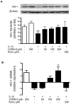
Figure 1. Effect of CORM-2 on HO-1 protein (A) and mRNA expression (B) in OA synoviocytes.
Cells were stimulated with IL-1β (10 ng/ml) for 24 h (A) and 16 h (B) in the presence or absence of CORM-2 (50, 100, 200 µM) or RuCl3 (200 µM). Protein expression was determined in cell lysates by Western blotting and mRNA levels were determined by real-time PCR. Relative expression of HO-1 and β-actin protein bands was calculated after densitometric analysis. Data are expressed as mean±S.E.M. Samples from 4 patients were used. ++P<0.01 with respect to nonstimulated cells. **P<0.01 with respect to IL-1β.
Figure 2
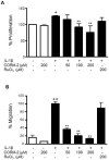
Figure 2. Effect of CORM-2 on synoviocyte proliferation and migration rate.
Cells were stimulated with IL-1β (10 ng/ml) for 24 h in the presence or absence of CORM-2 (50, 100, 200 µM) or RuCl3 (200 µM). (A) Cell proliferation was determined by the MTT assay. (B) Transwell chambers were kept in culture for 24 h. Upper compartment was detached and cells migrated to the lower side were counted in 6–8 microscopic power fields. Data (A) are expressed as mean±S.E.M. of % proliferating cells, considering 100% the proliferation induced in basal conditions whereas data (B) are expressed as % of cells migrated to the lower compartment, considering 100% the migration induced by IL-1β. Duplicate samples from 6 (A) and 4 (B) patients were used. +P<0.05, ++P<0.01 with respect to nonstimulated cells. **P<0.01 with respect to IL-1β.
Figure 3
Figure 3. Effect of CORM-2 on the levels of chemokines released into the medium by OA synoviocytes.
(A) IL-8, (B) CCL2 and (C) CCL20 protein levels. Cells were stimulated with IL-1β (10 ng/ml) for 24 h in the presence or absence of CORM-2 (50, 100, 200 µM) or RuCl3 (200 µM). Protein levels were determined in cell supernatants by ELISA. Data are expressed as mean±S.E.M. Duplicate samples from 6 patients were used. ++P<0.01 with respect to nonstimulated cells. *P<0.05 with respect to IL-1β.
Figure 4
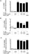
Figure 4. Effect of CORM-2 on chemokine mRNA levels in OA synoviocytes.
(A) IL-8, (B) CCL2 and (C) CCL20 mRNA levels. Cells were stimulated with IL-1β (10 ng/ml) for 16 h in the presence or absence of CORM-2 (100, 200 µM) or RuCl3 (200 µM). mRNA expression was determined by real-time PCR. Data are expressed as mean±S.E.M. Duplicate samples from 3 patients were used. ++P<0.01 with respect to nonstimulated cells. *P<0.05, **P<0.01 with respect to IL-1β.
Figure 5
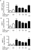
Figure 5. Effect of CORM-2 on MMP activity and MMP levels released into the culture medium in OA synoviocytes.
(A) MMP activity, (B) MMP-1 protein, (C) MMP-3 protein levels in the culture medium. Cells were stimulated with IL-1β (10 ng/ml) for 24 h in the presence or absence of CORM-2 (50, 100, 200 µM) or RuCl3 (200 µM). MMP activity was analyzed by fluorometric procedures in cell supernatants (A) and protein levels were determined by ELISA in cell supernatants (B–C), Data are expressed as mean±S.E.M. Duplicate samples from 6 patients were used. ++P<0.01 with respect to nonstimulated cells. *P<0.05 with respect to IL-1β.
Figure 6
Figure 6. Effect of CORM-2 on MMP mRNA levels in OA synoviocytes.
(A) MMP-1, (B) MMP-3 mRNA levels. Cells were stimulated with IL-1β (10 ng/ml) for 16 h in the presence or absence of CORM-2 (100, 200 µM) or RuCl3 (200 µM). mRNA expression was measured by real-time PCR. Data are expressed as mean±S.E.M. Duplicate samples from 6 patients were used. ++P<0.01 with respect to nonstimulated cells. *P<0.05, **P<0.01 with respect to IL-1β.
Figure 7
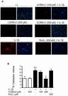
Figure 7. Effect of CORM-2 on oxidative stress levels in OA synoviocytes.
Cells were incubated with IL-1β (10 ng/ml) in the presence or absence of CORM-2 (100, 200 µM) or RuCl3 (200 µM) for 30 min. Oxidative stress was measured by the oxidation of dihydrorhodamine 123. Rhodamine positive cells were counted in 6–8 microscopic power fields. Data are expressed as % of oxidative stress. Left panels: oxidative stress, right panels: DAPI. Duplicate samples from 3 patients were used. +P<0.05 with respect to nonstimulated cells. *P<0.05 with respect to IL-1β. Original magnification: x200.
Figure 8
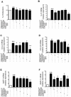
Figure 8. Effects of specific inhibitors of MAPK, Akt and NF-κB on IL-1β induced cell proliferation and chemokine and MMP mRNA expression in OA synoviocytes.
(A) Cell proliferation, (B) IL-8, (C) CCL2, (D) CCL20, (E) MMP-1, (F) MMP-3 mRNA expression. Cells were treated with UO126 (10 µM), SP600125 (25 µM), SB202190 (10 µM), MK2206 (20 µM) or PS1145 (10 µM), specific inhibitors of MEK/ERK1/2, JNK1/2, p38, Akt and IκB kinase, respectively, 1 h before adding IL-1β for a total incubation time of 24 h (A) or 16 h (B–F). Cell proliferation was determined by the MTT method whereas mRNA expression was analyzed by real-time PCR. Data are expressed as mean±S.E.M. Duplicate samples from 3 patients were used. +P<0.05, ++P<0.01 with respect to nonstimulated cells. *P<0.05, **P<0.01 with respect to IL-1β.
Figure 9
Figure 9. Effect of CORM-2 and IL-1β on Akt and MAPK phosphorylation in OA synoviocytes.
Cells were stimulated with IL-1β (10 ng/ml) for 5 min in the presence or absence of CORM-2 (100, 200 µM). Protein expression was determined in cell lysates by Western blotting using specific antibodies against phosphorylated or total proteins. Relative expression of phosphorylated and total protein bands was calculated after densitometric analysis. AU = arbitrary units. Data are expressed as mean±S.E.M. (samples from 3 patients). ++P<0.01 with respect to nonstimulated cells. *P<0.05, **P<0.01 with respect to IL-1β.
Figure 10
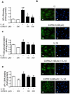
Figure 10. Effect of CORM-2 on NF-κB and AP-1 activation in OA synoviocytes.
(A) NF-κB binding to DNA, (B) NF-κB p65 translocation (left panels: p65, right panels: DAPI), (C) IκBα phosphorylation and (D) AP-1 binding to DNA. Cells were stimulated with IL-1β (10 ng/ml) for 30 min (C) or 1 h (A, B, D) with IL-1β (10 ng/ml) in the presence or absence of CORM-2 (100, 200 µM). IκBα phosphorylation and NF-κB/AP-1 binding to DNA were determined by ELISA in cytosolic and nuclear fractions respectively, whereas p65 translocation was analyzed by immunofluorescence. Original magnification x400. Data are expressed as mean±S.E.M. of independent cultures with cells from 3 different donors. AU = arbitrary units. +P<0.05, ++P<0.01 with respect to nonstimulated cells. *P<0.05, **P<0.01 with respect to IL-1β.

References

    1. Schedel J, Wenglen C, Distler O, Muller-Ladner U, Scholmerich J, et al. Differential adherence of osteoarthritis and rheumatoid arthritis synovial fibroblasts to cartilage and bone matrix proteins and its implication for osteoarthritis pathogenesis. Scand J Immunol. 2004;60:514–523. - PubMed
    1. Pelletier JP, Martel-Pelletier J, Abramson SB. Osteoarthritis, an inflammatory disease: potential implication for the selection of new therapeutic targets. Arthritis Rheum. 2001;44:1237–1247. - PubMed
    1. Benito MJ, Veale DJ, FitzGerald O, van den Berg WB, Bresnihan B. Synovial tissue inflammation in early and late osteoarthritis. Ann Rheum Dis. 2005;64:1263–1267. - PMC - PubMed
    1. Chevalier X. Upregulation of enzymatic activity by interleukin-1 in osteoarthritis. Biomed Pharmacother. 1997;51:58–62. - PubMed
    1. Schneider N, Mouithys-Mickalad AL, Lejeune JP, Deby-Dupont GP, Hoebeke M, et al. Synoviocytes, not chondrocytes, release free radicals after cycles of anoxia/re-oxygenation. Biochem Biophys Res Commun. 2005;334:669–673. - PubMed

Publication types

MeSH terms