. 2011 Oct 5;101(7):1720-9.
doi: 10.1016/j.bpj.2011.08.024.

Chain collapse of an amyloidogenic intrinsically disordered protein

Affiliations

Chain collapse of an amyloidogenic intrinsically disordered protein

Neha Jain et al. Biophys J. .

Abstract

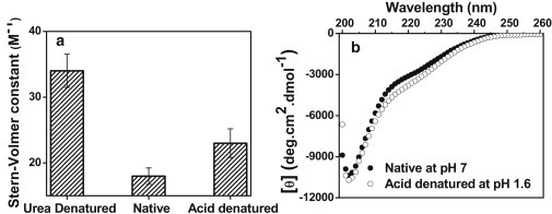
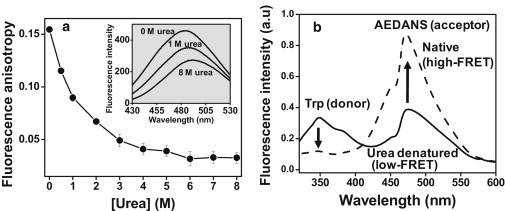
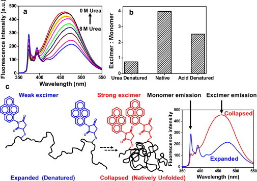
Natively unfolded or intrinsically disordered proteins (IDPs) are under intense scrutiny due to their involvement in both normal biological functions and abnormal protein misfolding disorders. Polypeptide chain collapse of amyloidogenic IDPs is believed to play a key role in protein misfolding, oligomerization, and aggregation leading to amyloid fibril formation, which is implicated in a number of human diseases. In this work, we used bovine κ-casein, which serves as an archetypal model protein for amyloidogenic IDPs. Using a variety of biophysical tools involving both prediction and spectroscopic techniques, we first established that monomeric κ-casein adopts a collapsed premolten-globule-like conformational ensemble under physiological conditions. Our time-resolved fluorescence and light-scattering data indicate a change in the mean hydrodynamic radius from ∼4.6 nm to ∼1.9 nm upon chain collapse. We then took the advantage of two cysteines separated by 77 amino-acid residues and covalently labeled them using thiol-reactive pyrene maleimide. This dual-labeled protein demonstrated a strong excimer formation upon renaturation from urea- and acid-denatured states under both equilibrium and kinetic conditions, providing compelling evidence of polypeptide chain collapse under physiological conditions. The implication of the IDP chain collapse in protein aggregation and amyloid formation is also discussed.


Figures

Figure 1
Figure 1
(a) Amino acid sequence of κ-casein. Trp76 and two Cys are shown with asterisk and underscoring, respectively. (b) Trp fluorescence spectra of 20 μM κ-casein under urea-denatured (dashed line) and native conditions (solid line). (c) Increase in Trp fluorescence anisotropy of κ-casein from the urea-denatured to the native condition.
Figure 2
Figure 2
(a) Stern-Volmer quenching constant of Trp of 20 μM κ-casein with acrylamide quencher under different conditions. For Stern-Volmer plots, see Fig. S3. (b) Far-UV CD spectra of 10 μM κ-casein under native (solid circles) and acid-denatured forms (open circles). Under the native condition, [θ]222 and [θ]200 were −2880 and −8880 deg·cm2 dmol−1, respectively.
Figure 3
Figure 3
(a) Increase in AEDANS fluorescence anisotropy upon refolding of fluorescently labeled 10-μM κ-casein from the urea-denatured state. (Inset) AEDANS fluorescence spectra as a function of denaturant concentration. (b) FRET between Trp (donor) and AEDANS (acceptor) under denatured and native conditions.
Figure 4
Figure 4
Picosecond time-resolved fluorescence anisotropy decays (r(t)) of Trp in denatured (a) and native (b) κ-casein. The solid line is the biexponential fit (Eq. 4). The recovered rotational correlation times, with associated amplitudes, are ϕfast = 0.2 ± 0.1 ns, βfast = 0.53 ± 0.02, ϕslow = 2.7 ± 0.8, βslow = 0.47 ± 0.02 for denatured κ-casein (a) and ϕfast = 0.2 ± 0.1 ns, βfast = 0.65 ± 0.03, ϕslow = 7.1 ± 0.6 ns, and βslow = 0.35 ± 0.03 for native κ-casein (b).
Figure 5
Figure 5
(a) Pyrene excimer formation of dual-labeled 10-μM κ-casein upon renaturation from urea. (b) Excimer/monomer ratio of pyrene under different conditions estimated from the fluorescence spectra shown in a. (c) A schematic of excimer formation upon polypeptide chain collapse.
Figure 6
Figure 6
(a) Trp fluorescence spectra of 20-μM κ-casein under acid-denatured and native conditions (b) Stopped-flow kinetics of Trp fluorescence upon acid renaturation of κ-casein from pH 1.6 to pH 7.0. An optical filter (320-nm long-pass) was used to collect total Trp fluorescence. The baseline is shown in gray. (c) Pyrene emission spectra of dual-labeled 10-μM κ-casein under acid-denatured and native conditions. (d) Stopped-flow kinetics of pyrene excimer formation upon acid renaturation. The optical filter (395-nm long-pass) used to selectively collect excimer fluorescence in the stopped-flow experiment is shown in c by a dotted line.

References

    1. Uversky V.N., Dunker A.K. Understanding protein non-folding. Biochim. Biophys. Acta. 2010;1804:1231–1264. - PMC - PubMed
    1. Dunker A.K., Lawson J.D., Obradovic Z. Intrinsically disordered protein. J. Mol. Graph. Model. 2001;19:26–59. - PubMed
    1. Tompa P. CRC Press; Boca Raton, FL: 2010. Structure and Function of Intrinsically Disordered Proteins.
    1. Uversky V.N. Intrinsically disordered proteins and their environment: effects of strong denaturants, temperature, pH, counter ions, membranes, binding partners, osmolytes, and macromolecular crowding. Protein J. 2009;28:305–325. - PubMed
    1. Radivojac P., Iakoucheva L.M., Dunker A.K. Intrinsic disorder and functional proteomics. Biophys. J. 2007;92:1439–1456. - PMC - PubMed

Publication types