

High-throughput analysis of concentration-dependent antibody self-association
- PMID: 21961601
- PMCID: PMC3183799
- DOI: 10.1016/j.bpj.2011.08.036
High-throughput analysis of concentration-dependent antibody self-association
Abstract
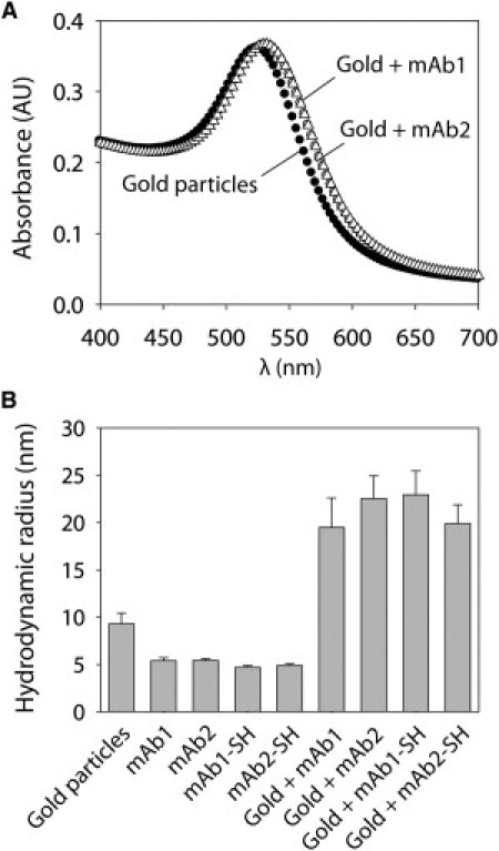
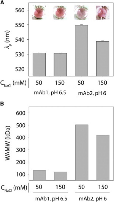
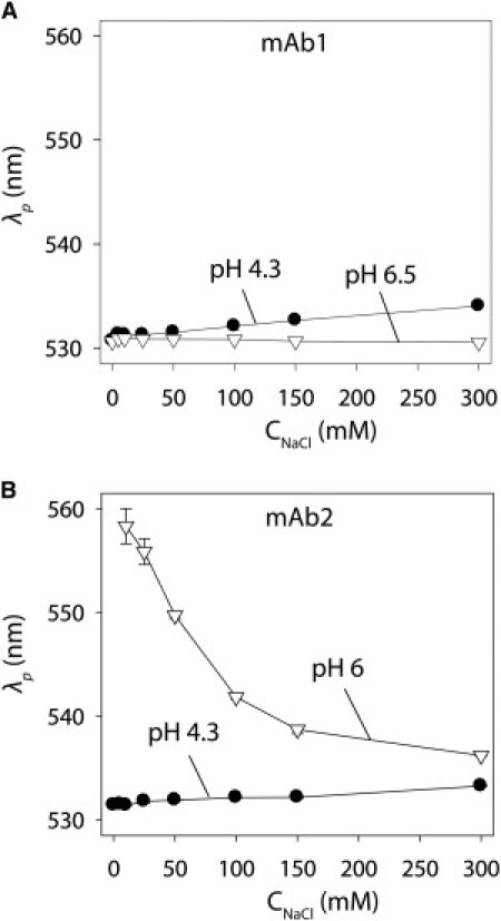
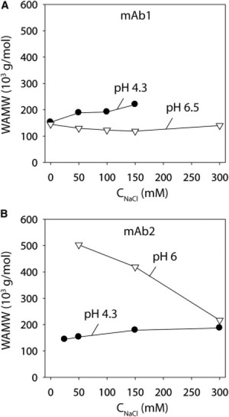
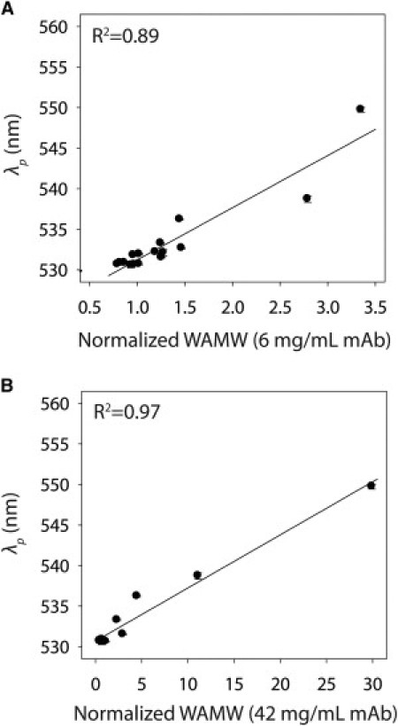
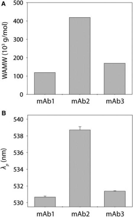
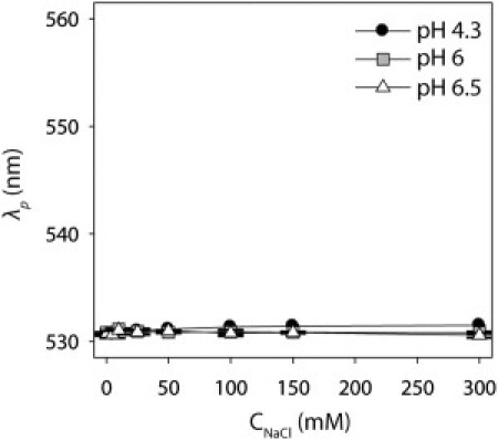
Monoclonal antibodies are typically monomeric and nonviscous at low concentrations, yet they display highly variable associative and viscous behavior at elevated concentrations. Although measurements of antibody self-association are critical for understanding this complex behavior, traditional biophysical methods are not capable of characterizing such concentration-dependent self-association in a high-throughput manner. Here we describe a nanoparticle-based method, termed self-interaction nanoparticle spectroscopy, that is capable of rapidly measuring concentration-dependent self-interactions for three human monoclonal antibodies with unique solution behaviors. We demonstrate that gold nanoparticles conjugated with antibodies at low protein concentrations (<40 μg/mL) display self-association behavior (as measured by the interparticle distance-dependent plasmon wavelength) that is well correlated with static light-scattering measurements obtained at three orders of magnitude higher antibody concentrations. Using this methodology, we find that the antibodies display a complex pH-dependent self-association behavior that is strongly influenced by the solution ionic strength. Importantly, we find that a polyclonal human antibody is nonassociative for all solution conditions evaluated in this work, suggesting that antibody self-association is more specific than previously realized. We expect that our findings will guide rational manipulation of antibody phase behavior, and enable studies that elucidate sequence and structural determinants of antibody self-association.
Copyright © 2011 Biophysical Society. Published by Elsevier Inc. All rights reserved.
Figures
References
-
- Voter W.A., Erickson H.P. The kinetics of microtubule assembly. Evidence for a two-stage nucleation mechanism. J. Biol. Chem. 1984;259:10430–10438. - PubMed
-
- Pollard T.D., Borisy G.G. Cellular motility driven by assembly and disassembly of actin filaments. Cell. 2003;112:453–465. - PubMed
-
- Chiti F., Dobson C.M. Protein misfolding, functional amyloid, and human disease. Annu. Rev. Biochem. 2006;75:333–366. - PubMed
-
- Shire S.J., Shahrokh Z., Liu J. Challenges in the development of high protein concentration formulations. J. Pharm. Sci. 2004;93:1390–1402. - PubMed
Publication types
MeSH terms
Substances
LinkOut - more resources
Full Text Sources
Other Literature Sources
Molecular Biology Databases
