

Characterization of enhanced monovalent and bivalent thrombin DNA aptamer binding using single molecule force spectroscopy
- PMID: 21961605
- PMCID: PMC3183820
- DOI: 10.1016/j.bpj.2011.07.054
Characterization of enhanced monovalent and bivalent thrombin DNA aptamer binding using single molecule force spectroscopy
Abstract
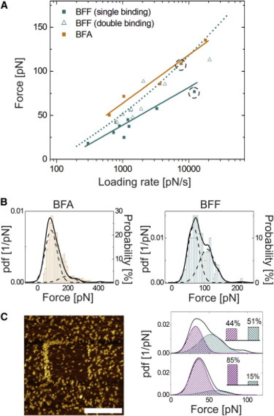
Thrombin aptamer binding strength and stability is dependent on sterical parameters when used for atomic force microscopy sensing applications. Sterical improvements on the linker chemistry were developed for high-affinity binding. For this we applied single molecule force spectroscopy using two enhanced biotinylated thrombin aptamers, BFF and BFA immobilized on the atomic force microscopy tip via streptavidin. BFF is a dimer composed of two single-stranded aptamers (aptabody) connected to each other by a complementary sequence close to the biotinylated end. In contrast, BFA consists of a single DNA strand and a complementary strand in the supporting biotinylated part. By varying the pulling velocity in force-distance cycles the formed thrombin-aptamer complexes were ruptured at different force loadings allowing determination of the energy landscape. As a result, BFA aptamer showed a higher binding force at the investigated loading rates and a significantly lower dissociation rate constant, k(off), compared to BFF. Moreover, the potential of the aptabody BFF to form a bivalent complex could clearly be demonstrated.
Copyright © 2011 Biophysical Society. Published by Elsevier Inc. All rights reserved.
Figures


Similar articles
-
Surface plasmon resonance spectroscopy study of interfacial binding of thrombin to antithrombin DNA aptamers.J Colloid Interface Sci. 2007 Nov 1;315(1):99-106. doi: 10.1016/j.jcis.2007.06.040. Epub 2007 Aug 8. J Colloid Interface Sci. 2007. PMID: 17689549
-
Investigation of the interaction between a bivalent aptamer and thrombin by AFM.Langmuir. 2012 Jan 10;28(1):707-13. doi: 10.1021/la203954x. Epub 2011 Dec 2. Langmuir. 2012. PMID: 22103891
-
Development of a novel aptamer-based sensing system using atomic force microscopy.J Biosci Bioeng. 2011 Nov;112(5):511-4. doi: 10.1016/j.jbiosc.2011.07.008. Epub 2011 Aug 6. J Biosci Bioeng. 2011. PMID: 21821470
-
Energy landscape of aptamer/protein complexes studied by single-molecule force spectroscopy.Chem Asian J. 2007 Feb 5;2(2):284-9. doi: 10.1002/asia.200600230. Chem Asian J. 2007. PMID: 17441163
-
Aptamer binding assays for proteins: the thrombin example--a review.Anal Chim Acta. 2014 Jul 21;837:1-15. doi: 10.1016/j.aca.2014.04.055. Epub 2014 May 2. Anal Chim Acta. 2014. PMID: 25000852 Review.
Cited by
-
Quantifying biomolecular hydrophobicity: Single molecule force spectroscopy of class II hydrophobins.J Biol Chem. 2021 Jan-Jun;296:100728. doi: 10.1016/j.jbc.2021.100728. Epub 2021 Apr 30. J Biol Chem. 2021. PMID: 33933454 Free PMC article.
-
High-Yield Characterization of Single Molecule Interactions with DeepTipTM Atomic Force Microscopy Probes.Molecules. 2022 Dec 27;28(1):226. doi: 10.3390/molecules28010226. Molecules. 2022. PMID: 36615422 Free PMC article.
-
Atomic force microscopy fishing and mass spectrometry identification of gp120 on immobilized aptamers.Int J Nanomedicine. 2014 Oct 3;9:4659-70. doi: 10.2147/IJN.S66946. eCollection 2014. Int J Nanomedicine. 2014. PMID: 25336946 Free PMC article.
-
[Application of atomic force microscopy-based single molecule force spectroscopy in G-quadruplex studies].Nan Fang Yi Ke Da Xue Xue Bao. 2018 Aug 30;38(9):1107-1114. doi: 10.12122/j.issn.1673-4254.2018.09.14. Nan Fang Yi Ke Da Xue Xue Bao. 2018. PMID: 30377115 Free PMC article. Review. Chinese.
References
-
- Ellington A.D., Szostak J.W. In vitro selection of RNA molecules that bind specific ligands. Nature. 1990;346:818–822. - PubMed
-
- Hianik T., Wang J. Electrochemical aptasensors-recent achievements and perspectives. Electroanalysis. 2009;21:1223–1235.
-
- Farokhzad O.C., Jon S., Langer R. Nanoparticle-aptamer bioconjugates: a new approach for targeting prostate cancer cells. Cancer Res. 2004;64:7668–7672. - PubMed
-
- Holland C.A., Henry A.T., Church F.C. Effect of oligodeoxynucleotide thrombin aptamer on thrombin inhibition by heparin cofactor II and antithrombin. FEBS Lett. 2000;484:87–91. - PubMed
-
- Bock L.C., Griffin L.C., Toole J.J. Selection of single-stranded DNA molecules that bind and inhibit human thrombin. Nature. 1992;355:564–566. - PubMed
Publication types
MeSH terms
Substances
LinkOut - more resources
Full Text Sources
Molecular Biology Databases
Research Materials
