

Vulvar squamous cell carcinoma with sarcoma-like stroma: a case report and review of the literature
- PMID: 21961623
- PMCID: PMC3192728
- DOI: 10.1186/1746-1596-6-95
Vulvar squamous cell carcinoma with sarcoma-like stroma: a case report and review of the literature
Abstract
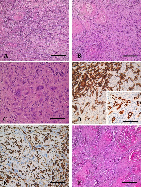
Vulvar squamous cell carcinoma with sarcoma-like stroma represents an extremely rare histological entity showing the co-existence of both epithelial and mesenchymal features: these tumors, firstly described in the skin by Martin and Stewart in 1935 have been further described in other anatomic sites including oral cavity, larynx, breast, lung and oesophagus. The complexity of the histology, as well as its aggressive clinical behaviour makes the diagnosis and the exploitment of effective therapeutic approaches very difficult, so that no definitive guidelines for treatments are currently available. Here, we describe a case of advanced stage vulvar squamous cell carcinoma with sarcoma-like stroma showing an unfavourable prognosis despite the use of an aggressive multimodal approach. A revision of the currently published cases have been also provided.
Figures
Similar articles
-
The prognostic significance of micrometastases in node-negative squamous cell carcinoma of the vulva.Br J Cancer. 2005 Jan 31;92(2):222-4. doi: 10.1038/sj.bjc.6602343. Br J Cancer. 2005. PMID: 15655537 Free PMC article.
-
Cytokine, cell adhesion receptor, and tumor suppressor gene expression in vulvar squamous carcinoma: correlation with prominent fibromyxoid stromal response.Int J Gynecol Pathol. 1996 Oct;15(4):320-5. doi: 10.1097/00004347-199610000-00004. Int J Gynecol Pathol. 1996. PMID: 8886879
-
Squamous cell carcinoma with sarcomatoid features of the vulva: a case report and review of literature.Gynecol Oncol. 2006 Oct;103(1):363-7. doi: 10.1016/j.ygyno.2006.05.031. Epub 2006 Jun 30. Gynecol Oncol. 2006. PMID: 16814852
-
Adenoid squamous carcinoma (pseudoangiosarcomatous carcinoma) of the vulva: a rare but highly aggressive variant of squamous cell carcinoma-report of a case and review of the literature.Int J Gynecol Pathol. 2008 Apr;27(2):288-91. doi: 10.1097/PGP.0b013e3181569904. Int J Gynecol Pathol. 2008. PMID: 18317210 Review.
-
Pediatric vulvar malignancies: rare but important to know.Semin Diagn Pathol. 2021 Jan;38(1):99-109. doi: 10.1053/j.semdp.2020.09.001. Epub 2020 Sep 5. Semin Diagn Pathol. 2021. PMID: 32943238 Review.
Cited by
-
Spindle cell morphology is related to poor prognosis in vulvar squamous cell carcinoma.Br J Cancer. 2013 Oct 15;109(8):2259-65. doi: 10.1038/bjc.2013.563. Epub 2013 Sep 24. Br J Cancer. 2013. PMID: 24064972 Free PMC article.
-
Vulvar Sarcomatoid Squamous Cell Carcinoma: A Rare Entity.Indian Dermatol Online J. 2023 Oct 5;14(6):856-860. doi: 10.4103/idoj.idoj_696_22. eCollection 2023 Nov-Dec. Indian Dermatol Online J. 2023. PMID: 38099028 Free PMC article.
-
Well-differentiated squamous cell carcinoma of the clitoris: a rare case report.Pan Afr Med J. 2024 Aug 5;48:153. doi: 10.11604/pamj.2024.48.153.44531. eCollection 2024. Pan Afr Med J. 2024. PMID: 39619415 Free PMC article.
-
Vulvar "proximal-type" epithelioid sarcoma: report of a case and review of the literature.Diagn Pathol. 2013 Jul 25;8:122. doi: 10.1186/1746-1596-8-122. Diagn Pathol. 2013. PMID: 23886403 Free PMC article. Review.
-
Mesonephric adenocarcinoma with a sarcomatous component, a notable subtype of cervical carcinosarcoma: a case report and review of the literature.Diagn Pathol. 2013 May 7;8:74. doi: 10.1186/1746-1596-8-74. Diagn Pathol. 2013. PMID: 23651629 Free PMC article. Review.
References
-
- Martin HE, Stewart FW. Spindle cell epidermoid carcinoma. Am J Cancer. 1935;24:273–298.
-
- Vulva. AJCC Cancer Staging Manual. 6. New York, NY: Springer; 2002. American Joint Committee on Cancer; pp. 243–9.
Publication types
MeSH terms
LinkOut - more resources
Full Text Sources
Medical
